By Md. R. Islam, F. Islam, MH. Nafady, T B. Emran, Simona Cavalu &al
Breast cancer (BrCa) is considered a global public health concern ; it is the second most widely diagnosed cancer and a prominent cause of mortality in women. As a result, to reduce the number of BrCa-related mortality, effective BrCa therapies are necessary. Furthermore, people of certain races or ethnicities are more likely to develop BrCa. African American women under the age of 40 are twice as likely to develop BrCA as white women of the same age. Females of American, African, and Hispanic heritage can be identified with aggressive and severe types of BrCa. Copyright Md.R. Islam, F. Islam, MH Nafady, Simona Cavalu & al.
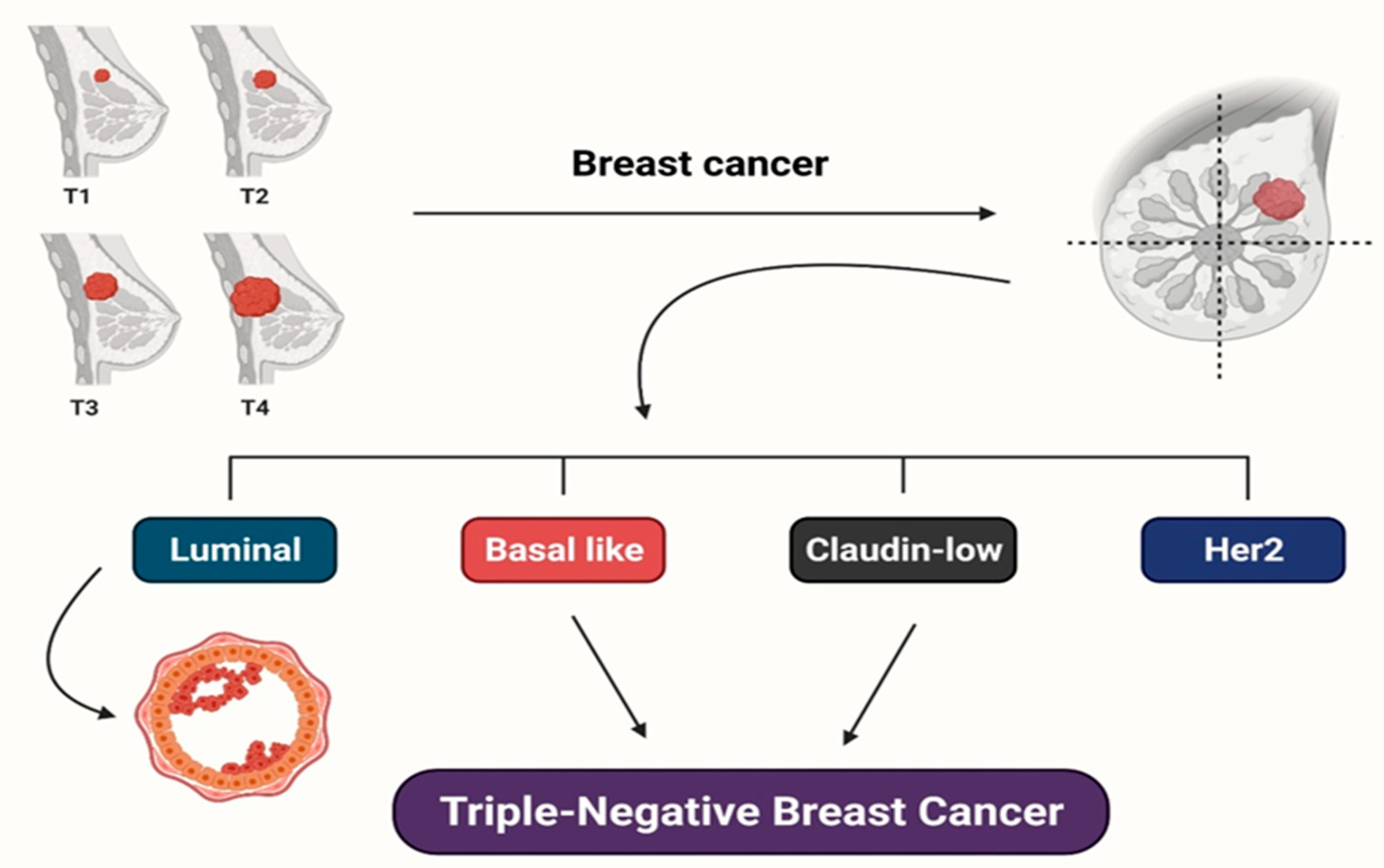
Overall survival (for breast cancer subtypes) and relapse-free survival (for TNBC subtypes) are used to distinguish the subtypes. Copyright Md.R. Islam, F. Islam, MH Nafady,
Simona Cavalu & al.
Risk Factors for Breast Cancer. Despite the urgent need for effective and novel therapies, there is considerable concern in identifying BrCa risk factors and improving chemo-preventive and lifestyle adjustment actions that can help decrease the impact of the disease. Although BRCA1 and BRCA2, as tumor-suppressor proteins, signify less than 10% of cases; their
discovery has overwhelmingly influenced patient treatment. Other risk factors linked with ER-positive BrCa progress, for example early menarche, early thelarche, and first pregnancy at a later age, are less well characterized and may also be linked to increased estrogen exposure. Obesity and metabolic syndrome, additionally, have been recently established as significant BrCa risk factors, a link that is particularly noteworthy considering the present obesity epidemic. Increased influence of adipokines and inflammatory cytokines, as well as increases in circulating insulin and insulin-like growth factors, local estrogen synthesis in adipose tissue, and the impact of circulating insulin and insulin-like growth factors, are all thought to play a role in disease development. Copyright Md.R. Islam, F. Islam, MH Nafady, Simona Cavalu & al.
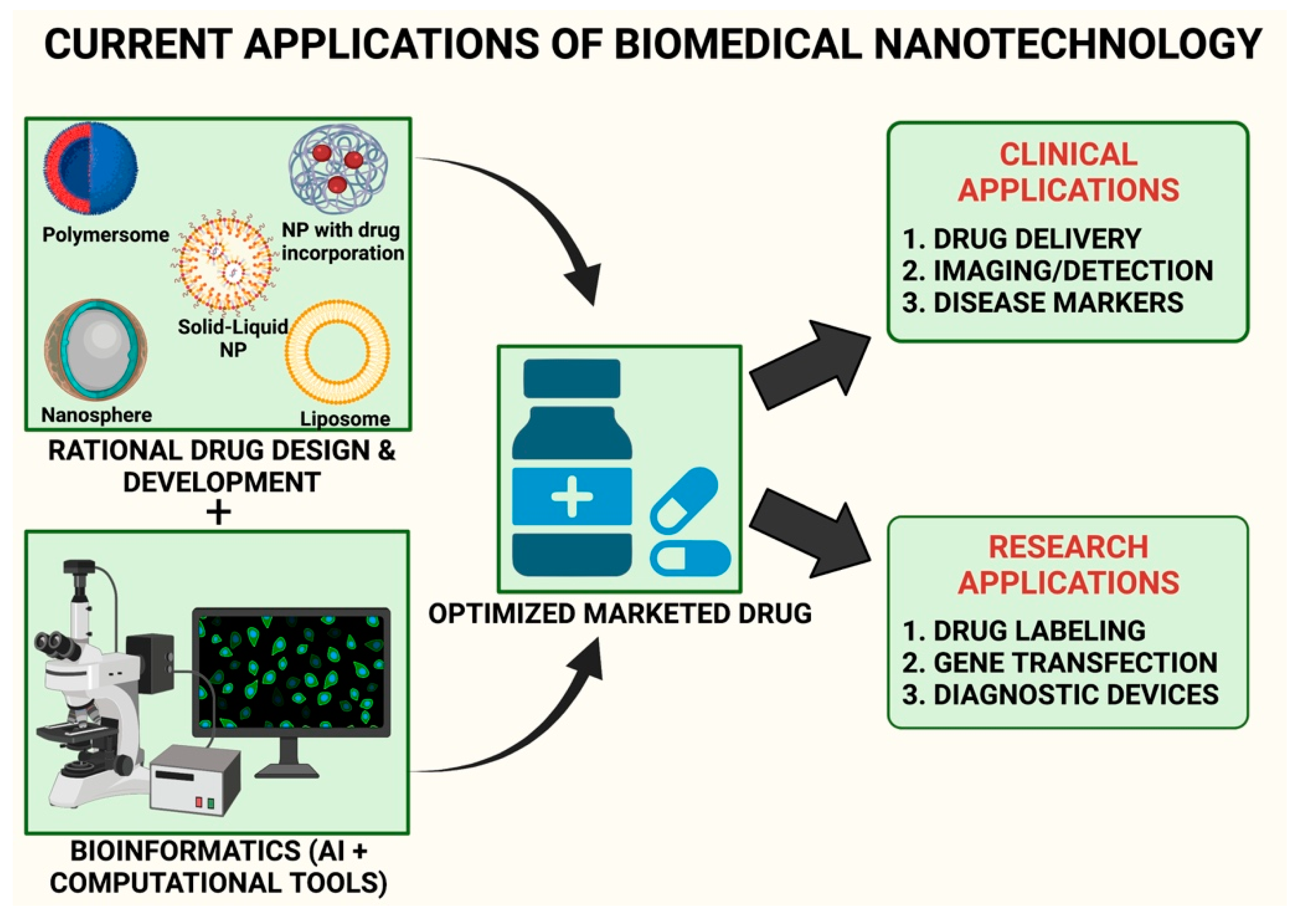
Natural Compounds against Breast Cancer: Quercetin, Tetrandrine, Thymoquinone, Resveratrol, Honokiol, Garcinol, Biochanin A, Lycopene, Shikonin, Sulforaphane, Echinacea, Garlic, Turmeric, Burdock, Carotenoids, Green Tea.
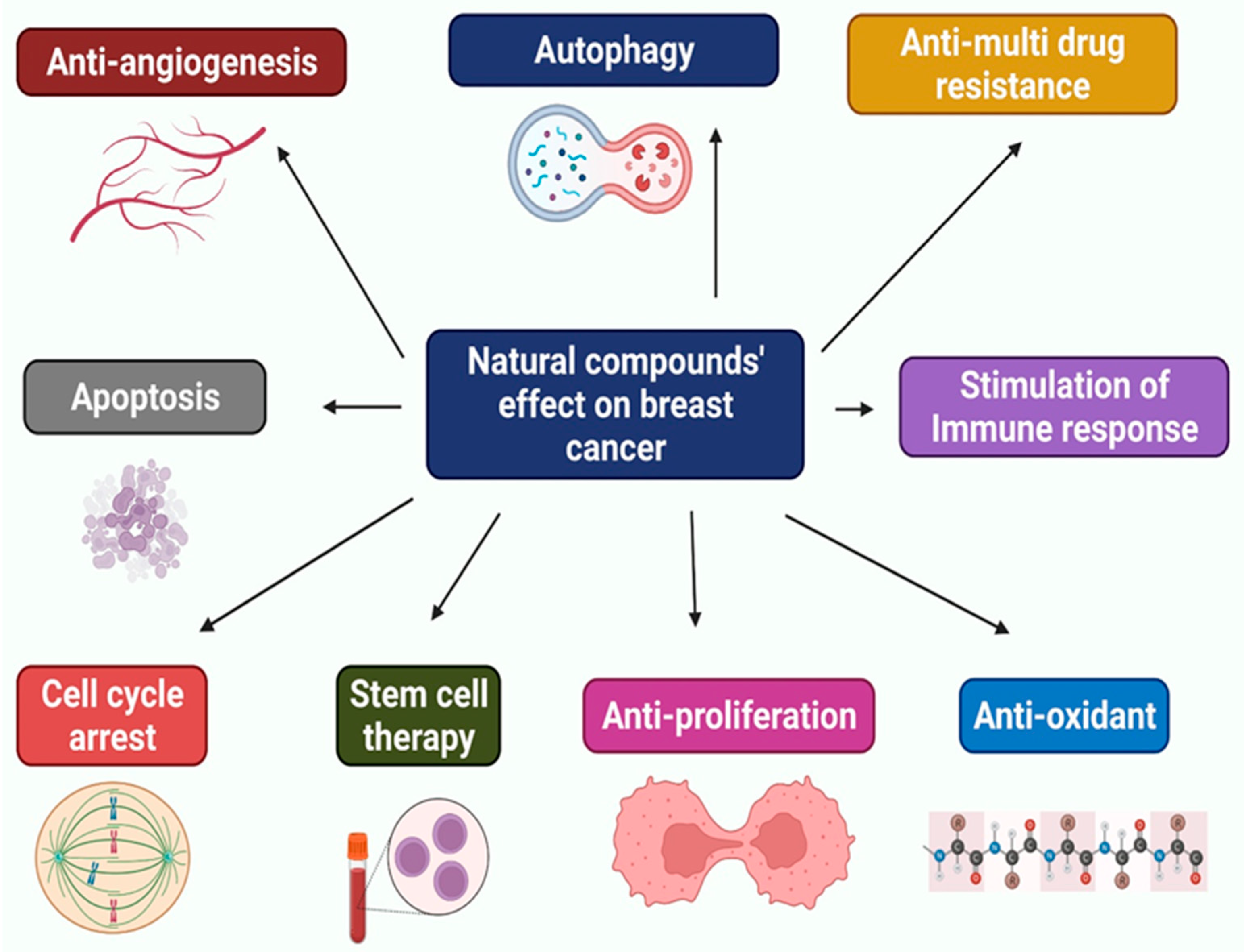
Natural substances have been shown through multiple investigations
to decrease carcinogenesis and reverse cancer growth by triggering apoptosis and cell cycle arrest. They impact tumor cells by interfering with cell death pathways such as extrinsic and intrinsic apoptosis and autophagy. These compounds inhibit cancer cell proliferation through these processes while causing minimal harm to normal cells. Natural compounds are currently being explored in clinical practice because of their anticancer
and apoptotic effects and low toxicity. Many of these substances will likely be used to treat BrCa as they have previously been found to have significant effects against various illnesses]. Finally, the natural compounds mentioned are just a fraction of the many chemicals that have been revealed to have anti-BrCa properties. Through the potential of these compounds, researchers are getting closer to finding a cure for BrCa. These compounds
have the potential to lower BrCa-related mortality and help people live longer across the world. Therefore, natural substances should continue to be investigated as an option for BrCa therapy. Copyright Md.R. Islam, F. Islam, MH Nafady, Simona Cavalu & al.
Full text available at https://www.mdpi.com/1420-3049/27/7/2165