By P. M. Reil, T. T. Maghiar, N. Vîlceanu, A. Pascalau, Claudia Teodora Judea Pusta, F. Marcu , Simona Cavalu and Ovidiu Pop
The aim of this our study was to determine any significance of the mCD14 and sCD14 levels in the septic cardiomyopathy, and to evaluate the correlation with lipopolysaccharide-binding protein (LBP) by means of immuno-histological examinations.
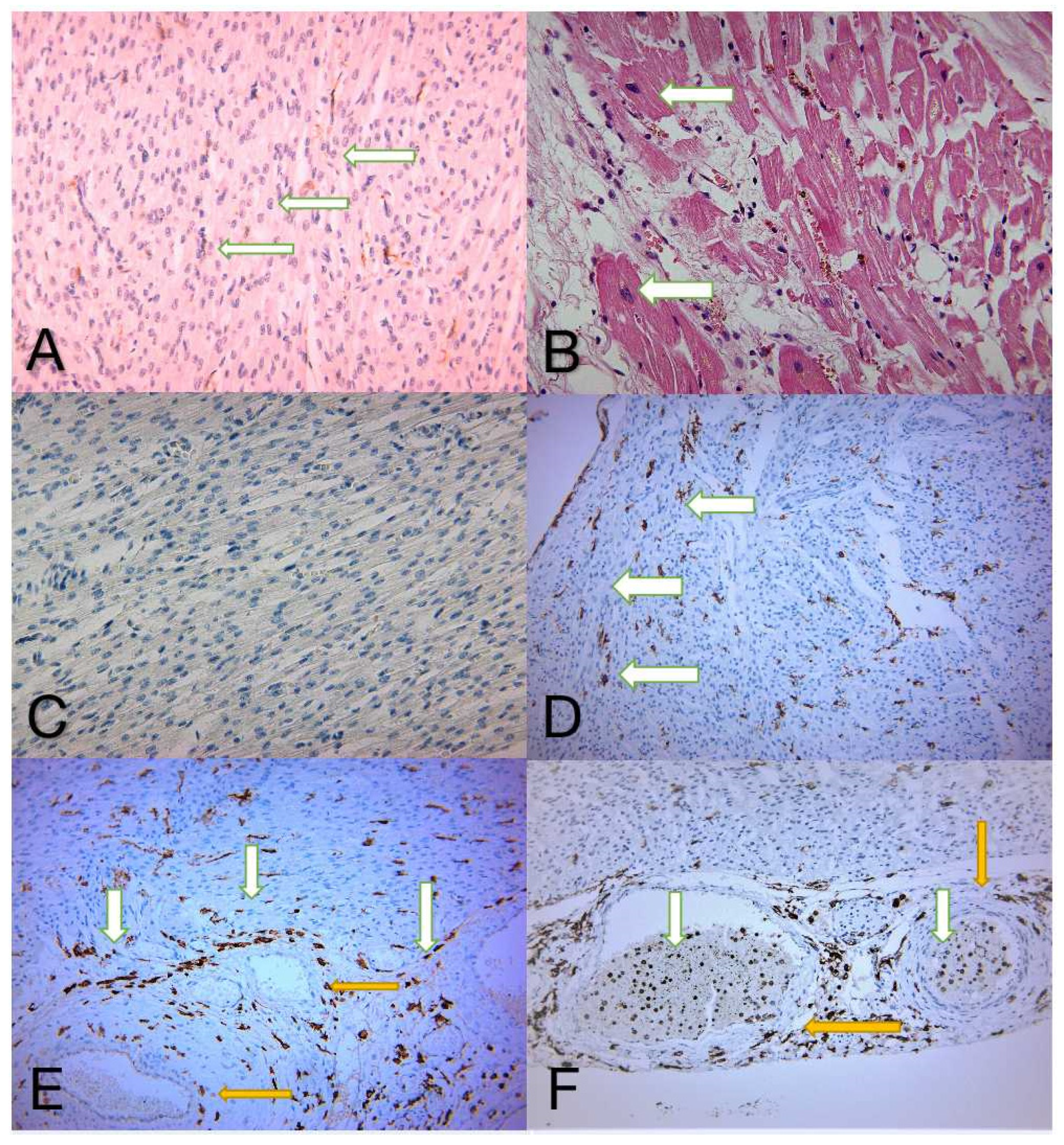
The study showed: 1) a positive association between markedly increased values of CD14 and an adverse patient evolution; 2) significant increase in the level of membranous and soluble CD14 surface protein in the study group; 3) tendency for higher values in relation to older patients, but without statistical significance; 4) no statistically relevant difference regarding the patient’s gender, provenance, or infection site; 5) a positive association between cellular expression (membranous, mCD14) and intravascular (soluble, sCD14) levels; 6) CD14 plays a double role: (a) as a component of the innate immune system, a pattern recognition receptor (PRR) proposed to bind conserved molecular structures on microbes (pathogen-associated molecular patterns, PAMPs, e.g., LPS); (b) as an active agent involved in the apoptotic cell cleaning process. Apoptotic cell-associated molecular patterns (ACAMPs) interfere with PRR and LPS-like
structures, as revealed on apoptotic cells. (Copyright P. M. Reil, T. T. Maghiar, N. Vîlceanu, A. Pascalau, Claudia Teodora Judea Pusta, F. Marcu , Ovidiu Pop and Simona Cavalu)
We suggest that a large amount of sCD14 detected in the myocardial tissue will activate the mCD14–TRL4–LBP–LPS complex and further induce an inadequate immune response, resulting in severe heart damage. A higher amount of LPS will induce more significant heart damage. Identification of the presence of mCD14 and sCD14 in the myocardium tissue can allow the indubitable diagnosis of septic cardiomyopathy. (Copyright P. M. Reil, T. T. Maghiar, N. Vîlceanu, A. Pascalau, Claudia Teodora Judea Pusta, F. Marcu , Simona Cavalu and Ovidiu Pop)
Full text available at: https://www.mdpi.com/2075-4418/12/4/781/htm
