The purpose of our study was to investigate the structural modifications of collagen extracellular matrix of amniotic membrane upon interaction with two different antibiotics, frequently used in surgical and post- surgical procedure, respectively ciprofloxacin and gentamicin. SEM micrographs evidenced the ultrastructure features of dried amniotic membrane, with laminar structure, flexible, transparent, with no blood vesels or nerves. FTIR spectroscopy combined with deconvolution techniques was applied with the aim to determine the extent of denaturation upon treatment with different antibiotics. By spectral analysis, we concluded that gentamicin treatment is more favorable compared to ciprofloxacin, as the denaturation process is reflected by the lower sheet/turns ratio of the secondary structure composition.
Photographic image of amniotic membrane; (b) SEM micrograph recorded on the surface of amniotic membrane; (c) SEM cross-section image of amniotic membrane. Copyright Simona Cavalu et al.FTIR spectra of amniotic membrane: (a) air dried, natural; (b) after gentamicin treatment; (c) after ciprofloxacin treatment. Copyright Simona Cavalu et al.Quantitative analysis (percent) based on spectral deconvolution of amide I FTIR absorption band of natural amniotic membrane and threated with gentamicin and ciprofloxacin. Copyright Simona Cavalu et al.
The present study was devoted to structural analyses of amniotic membrane as a potential natural biomaterial for biomedical applications, including tissue regeneration. The basement membrane closely resembles to that of the conjunctiva and cornea, especially with regards to its collagen composition (collagen type IV, V and VI) in addition to fibronectin and laminin. SEM micrographs evidenced the ultrastructure features of dried amniotic membrane, with laminar structure, flexible, transparent, with no blood vessels or nerves. FTIR spectroscopy combined with deconvolution techniques was applied in order to determine the extent of amniotic membrane denaturation upon treatment with different antibiotics. By spectral analysis, we can conclude that the gentamicin treatment is more favorable compared to ciprofloxacin, as the denaturation process is reflected by the lower sheet/turns ratio.(Copyright Simona Cavalu et al.)
The aim of this study is to obtain “giant” liposomes by lipid film hydration using a preparation formula with two different phospholipids, phosphatidylcholine (PC) and phosphatidylserine (PS).Firstly, the macro- and microscopic characterization, total phenols content and antioxidant capacity of the plant Stellaria media (L.) Vill. were assessed. Then, Stellaria media (L.) Vill. extract was encapsulated in both formulations (PCE and PSE) and the liposomes were characterized according to their morphology, size distribution and Zeta potential using optical microscopy and dynamic light scattering. The encapsulation efficiency (EE%) was determined using the Folin–Ciocalteu method and the values of both formulations were compared. PC and PCE liposomes with a diameter between 712 and 1000 nm and PS and PSE liposomes with a diameter between 58 and 1000 nm were obtained. The values EE% of Stellaria media (L.) Vill. extract for PCE and PSE were 92.09% and 84.25%, respectively.
Preparation of the liposomes with Stellaria media (L.) Vill. extract and empty liposomes by lipid film hydration. Copyright F. Miere(Groza), Simona Cavalu et al.Macroscopic and microscopic characteristics of the plant Stellaria media (L.) Vill. (a) The aerial part of the plant Stellaria media (L.) Vill. Stelariae herba (personal photo), (b) flower of the plant Stellaria media (L.) Vill. (personal photo), (c) cross-section through the main stem (100×), (d) longitudinal section through the main stem (200×). Copyright F. Miere (Groza), Simona Cavalu et al.Microscopic images of the liposomes: (a) Liposomes with phosphatidylcholine (PC), (b) Liposomes with phosphatidylcholine with encapsulated Stellaria media (L.) Vill. extract (PCE), (c) Liposomes with phosphatidylserine (PS), (d) Liposomes with phosphatidylserine with encapsulated Stellaria media (L.) Vill. extract (PSE). The red arrows show the characteristic spherical shape of liposomes. Copyright F. Miere (Groza), Simona Cavalu et al.Diameter distribution for PC liposomes without encapsulated extract of Stellaria media (L.) Vill. (a) and PCE liposomes with encapsulated extract of Stellaria media (L.) Vill. (b). Copyright F. Miere (Groza), Simona Cavalu et al.
Both PC and PS liposomes and their homologues with encapsulated plant extract were “giant” multilamellar liposomes. In the case of PC and PCE liposomes, around 50–80% presented dimensions between 712 and 1000 nm, while more than 90% of PS and PSE liposomes were in the range of 58–1000 nm. The larger diameter of the PC and PCE liposomes confirmed that the type of phospholipids used in the preparation significantly influenced the size and electrical charge of the formulation. The phosphatidylserine-based formulations showed smaller diameters and a negative Zeta potential, meaning they had better stability compared to phosphatidylcholine-based ones. We also demonstrated a high inclusion percentage of the Stellaria media (L.) Vill. extract in both formulations—more than 90% for PCE and more than 80% for PSE. (Copyright F. Miere (Groza), Simona Cavalu et al.
Knowing the biological and pharmacological properties of propolis, the first goal of our study was to prepare and characterize a propolis nano-formulation (NPs) in order to be used for wound healing applications. The ability of propolis NPs to stimulate the migration of dermal fibroblasts in vitro was assessed by scratch test assay. The concentration of 200 μg/mL propolis NPs was found to have similar effect as the positive control. The second goal was to prepare a propolis-collagen membrane and to investigate the morphological and nanoindentation properties by AFM. The ultrastructure network of collagen fibrils was not affected by incorporation of propolis NPs, showing a nano-porous structure, favorable for soft tissue regeneration applications. Enzymatic degradation assay indicated a reduced degradation rate upon incorporation of propolis NPs in collagen matrix.
Ionotropic gelation method was applied for the preparation of propolis NPS. The nanoparticles were formed spontaneously due to ionic interaction between the protonated amine groups in chitosan and the negatively charged counter-ion TPP, being stabilized by Tween 80.
TEM image of propolis NPs; b) size distribution and c) EDX corresponding spectrum. Copyright Simona CavaluSpontaneous evolution of human fibroblasts in cell culture medium, monitored at different time intervals (6, 12, 24, 48, 96 and 110 hours) until the confluence was achieved (Phase contrast image, scale bar 50 μm). Copyright Simona CavaluFibroblasts migration monitored after different time intervals and wound closure under the treatment with propolis NPs at two different concentrations, compared to the positive and negative control. The initial area of the scratch (t=o) is represented by the red rectangle (Phase contrast image, scale bar 100 μm). Copyright Simona Cavalu.The percent of restored fibroblasts monolayer upon migration of the cells into the free area, monitored during 48 h (Statistical relevance p<0.05). Copyright Simona Cavalu.AFM images of neat collagen membrane (a,b) and collagen membrane with propolis NPs incorporated (c, d), in 3D and 2D configurations.
The tridimensional network of collagen fibrils is visible in both specimens (with or without propolis NPs incorporated) emphasizing the details of repetitive structure of the D-bands pattern of a single collagen fibril, with periodic gaps and grooves, in concordance with some previous published work [32, 33]. The periodicity of D-bands is less visible after propolis NPs incorporation. Moreover, after propolis NPs incorporation and freeze drying procedure, an obvious porous ultrastructure formation was noticed, as a result of fibers self-assembly.
Collagenase degradation test of neat collagen membrane and collagen-propolis NPs membrane (statistical relevance p<0.05). Copyright Simona Cavalu.
A collagen-based membrane was prepared and investigated by AFM in terms of morphological features and nanoindentation. The network of collagen fibrils was not affected by propolis NPs, showing a nano-porous structure, favorable for soft tissue regeneration applications. Enzymatic degradation assay indicated a reduced degradation rate upon incorporation of propolis NPs in collagen matrix. Corroborating the above mentioned results, we consider that modified-collagen membrane by adding propolis NPs in a controlled concentration, might represent a promising natural alternative to synthetic bandages for wound healing applications. Of course, further in vitro and in vivo tests are necessary to evaluate the biological performances of collagen-modified membranes, in terms of tissue adaptation and integration. (Simona Cavalu, PM Pasca, Digest Journal of Nanomaterials and Biostructures, Volume 16, Issue 3, Pages 929 – 938July-September 2021).
By Laslo, V., Pinzaru, S. C., Zaguła, G., Kluz, M., Vicas, S. I., & Simona Cavalu
Cadmium is a heavy metal, environmental pollutant and toxic for organisms. Lactic acid bacteria (LAB) and nanoparticles represent useful tools to remove heavy metals from different environments. The main goal of our study was to develop a novel experimental design for cadmium (Cd) disposal using L. casei or L. fermentum and exogenous selenium nanoparticles. The experimental design was developed in two steps. In the first step, bio-synthesis, morphological and chemical characterization of selenium nanoparticles (SeNPs) produced by L. casei was performed. In the next step, L. casei and L. fermentum were used to evidence their binding ability toward Cd, highlighting the role of SeNPs against Cd-induced toxicity. Homogenous, spherical shape SeNPs and sharp size distribution with maximum 200 nm were obtained via reduction route, using L. casei and Na 2 SeO 3 . Addition of Cd to LAB culture medium strongly inhibits both bacteria growth. SeNPs, in contrast to sodium selenite, clearly demonstrates the protective effect against Cd toxicity, as evidenced by dynamic growth curves of both bacteria. A better stimulation of endogenous SeNPs production was noticed for L. fermentum compared to L. casei . The efficiency of Cd bioabsorption by LAB was in the range 43.78% – 50.87%, the ratio of 1:2 LAB/SeNPs showing the best results for both bacteria. The synergic effect of biogenic SeNPs and LAB against Cd toxicity was demonstrated. The combination of FTIR (Fourier Transform Infrared), FT Raman (Fourier Transformed Raman) and ICPOES (Inductively Coupled Plasma Optical Emission Spectroscopy) techniques provides specific qualitative and quantitative information on the behavior of probiotic species with respect to SeNPs synthesis and Cd disposal. (Simona Cavalu et al. JOMS 1247, 2022, 131325).
Liquid crystals (LCs) are also known as mesophase, which means an intermediate state between conventional solids and isotropic liquid, being mostly classified into two types: thermo tropic LCs (phase transition as a function of temperature) and lyotropic LCs (phase transition as a function of concentration of amphiphiles). Important features like thermodynamic stability, improved solubility of hydrophobic drugs, improved bioavailability and controlled release pattern made them effective carriers for a variety of drugs and bioactive compounds. Due to these unique features, LCs possess wide applications in the field of pharmaceuticals and become an attractive choice of vehicle for in vivo drug delivery. This review paper aims on highlighting the concept of LCs, classification, preparation methods and characterization techniques, in the context of pharmaceutical applications along with its perspectives in drug delivery systems. (Rajni Bala, Rakesh K. Sindhu, Bharti Kaundle, Reecha Madaan, Simona Cavalu, JOMS 1245, 2021, 131117)
Simultaneous radiotherapy and hyperthermia yield synergistic improvement of therapeutic efficacy in cancer therapy. The study is focussed on sol-gel derived particles of 60SiO2•20Al2O3•10Fe2O3•10Dy2O3 (mol%) system functionalized with type I collagen. The addition to aluminosilicate matrix of Fe2O3•was thought for hyperthermia and of Dy2O3 for internal radiation therapy after neutron activation of dysprosium to 165Dy radioisotope. The collagen functionalization of these particles aimed to make them well accepted in the body and to contribute to destruction of reactive oxygen species, what has to be taken into account when nanoparticles are used for cancer therapy.
The XRD, FTIR and XPS investigations were carried out on Fe-Dy-aluminosilicate particles of average size around tens of nanometers, before and after their incubation in collagen solution in order to prove the protein functionalization particles and to assess the changes in secondary structure of protein following this process. The results show the anchoring of the protein on particles surface in a few nanometers thin layer as well as some relative changes of protein secondary structure. After interface processes with particles surface, the secondary structure of amide I in collagen consists overwhelmingly of α-helices and only about 12 % of β-turns structure (S. Simon, Simona Cavalu, D. Eniu, V. Simon, JOMS 1236, 2021,130341).
By Inna V. Fadeeva, Margarita A. Goldberg, Ilya I. Preobrazhensky, Georgy V. Mamin, Galina A. Davidova, Nadezhda V. Agafonova, Marco Fosca, Fabrizio Russo, Sergey M. Barinov, Simona Cavalu & Julietta V. Rau
Brushite cements based on powders of Zinc (Zn) (1.4 wt%) substituted tricalcium phosphate (β-TCP) and non-substituted β-TCP were prepared and investigated by SEM/XRD, MTT test and microbiology assay. Copyright J. Rau, Simona Cavalu et al.
Zinc (Zn) is an important biological trace element that plays a role in the normal growth and development of the skeleton. Its content in human bones (0.0126–0.0217 wt%) is about 28% of the total amount of Zn in the body (0.0030 wt% of Zn in tissues) [20]. The lack of Zn slows down the growth of the bone mass and has a negative influence on the bone metabolism [21]. On the other hand, Zn deficiency is a factor of risk for bone osteoporosis [22].
XRD spectra of: TCP-based cement (A) before and (B) after soaking in physiological solution and Zn-substituted TCP cement (C) before and (D) after soaking in physiological solution. Copyright J. Rau, Simona Cavalu et al.FTIR spectra of: A non-substituted and Zn-substituted TCP powders, B non-substituted and Zn-substituted TCP cements, and C non-substituted and Zn-substituted TCP cements after soaking in physiological solution. Copyright J. Rau, Simona Cavalu et al. Comparison of EPR spectra for Zn-TCP powder sample annealed at 900 °C before and after X-ray irradiation. Copyright J. Rau, Simona Cavalu et al.Bone Cements’ morphology, SEM images: A, B cement based on β-TCP; C, D cement based on Zn-β-TCP; E, F cement based on β-TCP after soaking in physiological solution; G, H cement based on Zn-β-TCP after soaking in physiological solution. Copyright J. Rau, Simona Cavalu et al.The inhibition of growth of: E. Coli for A Zn-TCP cement and B TCP cement; CE. faecium, and DP. aeruginosa for Zn-TCP cement (1) and TCP cement (2). The Zn-substituted TCP cement shows a stronger antibacterial activity, which is expressed in a larger bacterial zone of inhibition, with respect to the TCP cement. Copyright J. Rau, Simona Cavalu et al.
In this work, the cement based on the Zn-substituted β-TCP powder with a simplified preparation recipe and improved characteristics was developed. The setting time of cements was 8 min (the ratio of cement powder: hardening liquid = 3:1), which is optimal for preparation and application of the developed cements for bone defects during surgery. The Zn2+ content was selected to be 1.40 wt%. The pH of the cements reached 6.5 within 60 min after setting. After soaking in physiological solution for 60 days, the morphology and composition of cements changed. The final phases were DCPD and HA.The EPR measurements showed the presence of the trapped hydrogen and confirmed that annealing at 900 °C led to the significant reduction of carbonate impurities embedded into the β-TCP structure.The NCTC L929 fibroblast cell viability on the developed Zn-β-TCP cement was 10% higher compared to cement without Zn and possess antibacterial properties against E. coli, E. faecium, and P. aeruginosa.This finding confirms that the novel material could be a valid strategy for a range of biomedical application in humans. Therefore, it could offer promising potential for bone replacement and repair in moderate and non-load-bearing defects that are prone to infection in orthopedic and trauma setting.
Stages of wound healing and the implications of fibroblasts in this process. Copyright F. Groza, Simona Cavalu et al.
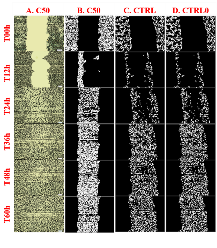
The main aim of our work was to highlight the in vitro healing potential of Stellaria media (L.) Vill. (SM) extract using the scratch assay on normal human dermal fibroblasts (NHDF). The ability to stimulate cell migration and proliferation under the influence of different concentrations of SM extract (range between 12.5 and 200 µg/mL) was determined compared to the control (untreated in vitro-simulated wound) and positive control (allantoin 50 µg/mL).Our findings demonstrate, for the first time in the literature, the wound-healing potential of SM extract on NHDF using the in vitro scratch method.
Effect of Stellaria media extract concentrations (between 12.5 µg/mL and 200 µg/mL) on viability of NHDF after 24 h. Copyright F. Miere, Simona Cavalu et al.The evolution over time (0, 12, 24, 36, and 48 h) of cell wound coverage at different concentrations of Stellaria media (L.) Vill extract (range 200 µg/mL–12.5 µg/mL) compared to positive control (CTRL)—treated with allantoin 50 µg/mL and untreated wound control (CTRL0). Fibroblasts marked in pink are those that have mobilized into the wound. The scale of the processed images is 100 µm. Copyright F. Groza, S. I. Vicas, Simona Cavalu et al.The evolution of the cell density parameter inside the wound depending on the applied treatment and time. Copyright F. Groza, Simona Cavalu et al.Evolution over time, of the average values of the widths (µm) of the wound, the areas (mm2) of the wound, of the normalized densities (%) of the cells inside the wound, and the Lp norm, for all treatments. C200—12.5 µg/mL represents the Stellaria media (L.) Vill extract concentrations applied, CTRL—positive control (allantoin—50 µg/mL), CTRL0—control (without treatment), where no treatment was applied. T00–T48 represents the time expressed in hours. Copyright F. Groza, Simona Cavalu et al.
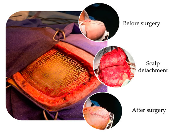
Titanium cranioplasty procedure in the case of a large defect. Copyright Simona Cavalu et al.
The Importance of Nano-Structured Surface on Titanium Implants. Titanium mesh or plates can be used in cranioplasty either alone or in conjunction with other synthetic materials, such as hydroxyapatite, calcium phosphate, and polyethylene.
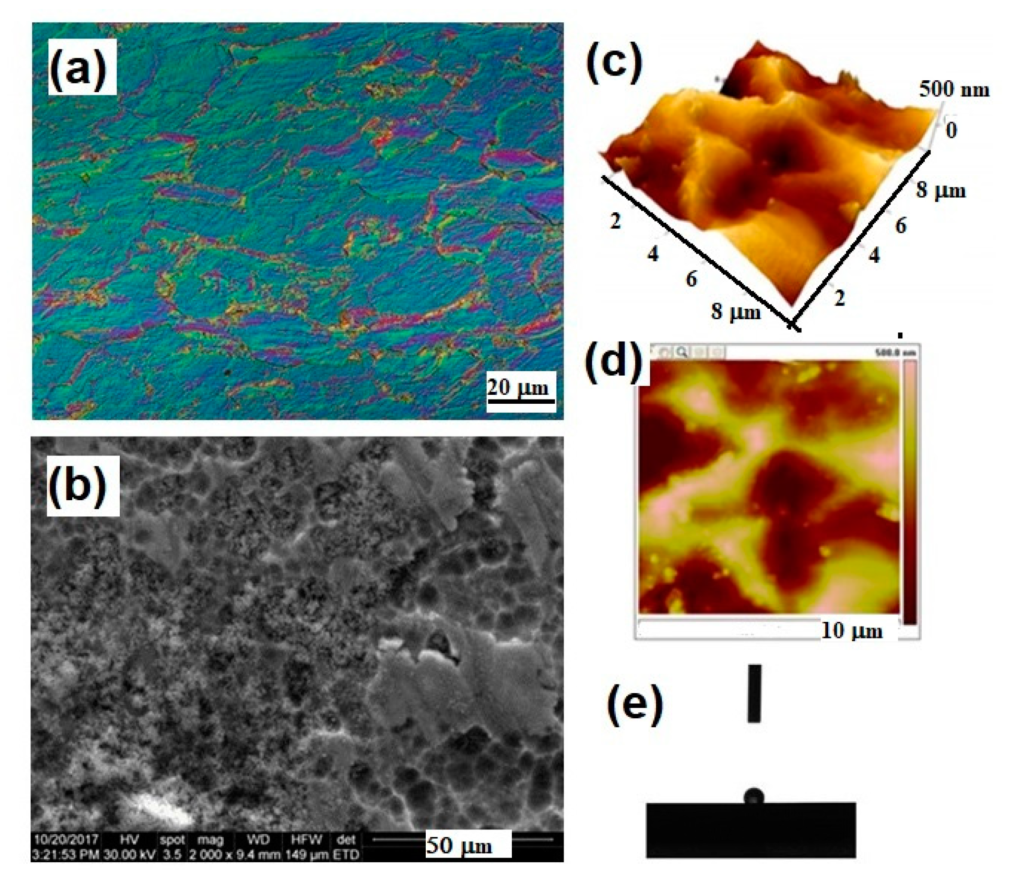
Surface properties of Ti mesh for cranioplasty evidenced by different microscopic techniques. Copyright Simona Cavalu et al.The main sequence of events occurring in vivo, during interaction between Ti surface and biological environment. Copyright Simona Cavalu et al.(a) The flow chart of SeNPs production via hydrothermal reaction using different saccharides as reducing agent; (b) TEM image of SeNPs used for the surface modifications of Ti mesh for cranioplasty, along with the surface morphology of the coating upon in situ SeNPs deposition and details of fibroblasts adhesion on the nanostructured Ti surface. Copyright Simona Cavalu et al.
This review emphasized the main reasons why titanium mesh is preferred for skull reconstructions along with the importance of developing innovative surface structures with a dual benefit in terms of improved osteointegration and enhanced antibacterial activity to reduce the risk of post-surgical infection, knowing that infections are the main complication in cranioplasty surgeries.
Biological effects of essential oils on the CNS through activation of various components of the brain. Copyright Simona Cavalu et al.
Since ancient times, essential oils (EOs) have been widely used and have been identified as therapeutic agents owing to their pharmacological and psychological properties. They were deemed to be physical, spiritual, and mental healing agents [1,2]. EOs are naturally occurring complex mixtures of volatile odor compounds synthesized as secondary metabolites by plants and are extracted through steam distillation, solvent extraction, maceration, cold press extraction, water distillation, and CO2 extraction. Novel methods that are more efficient and provide higher yields include supercritical fluid extraction, microwave-assisted extraction, and ultrasound [3]. Studies conducted on animals and humans have shown that EOs can produce a variety of CNS targeted pharmacological effects such as anxiolytic effect, neuroprotection, antidepressant effect, anticonvulsant effect, analgesic, and sedative effect, to name a few. As a result, EOs can be used as an adjuvant therapy to prevent and relieve symptoms associated with CNS-based disorders such as insomnia, depression, dementia, Alzheimer’s disease (AD), etc. As they are naturally occurring, they have the added benefit of being non-toxic and safe when utilized correctly at appropriate concentrations, which have been proven through research in the last ten years.
AuNPs as anticancer therapeutics. Copyright Simona Cavalu et al.Role of essential oils in acetylcholine release and degradation. Copyright Simona Cavalu et al.
Nanozymes can be controlled remotely via stimuli including heat, light, magnetic field, and ultrasound. Collectively, these all can be used to increase the therapeutic as well as diagnostic efficacy.
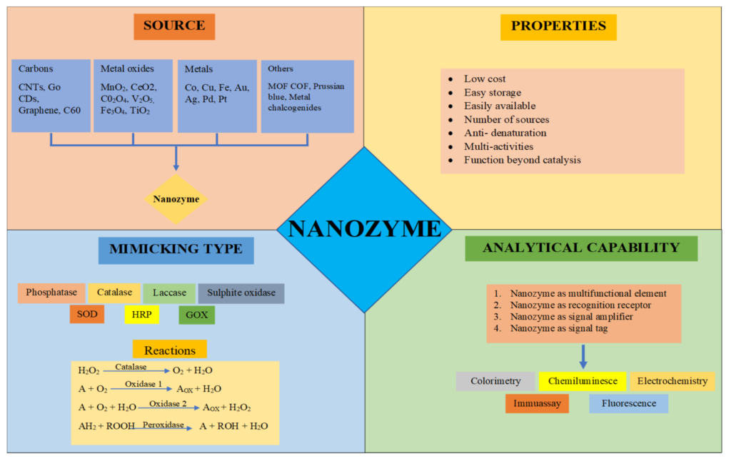
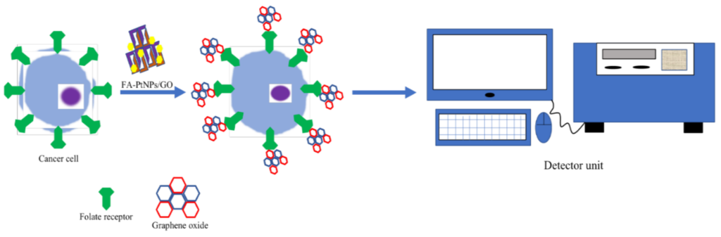
Sources, properties, mimicking types, and analytical capabilities of nanozymes. Copyright Simona Cavalu et al.Opportunities in the field of nanozymes. Copyright Simona Cavalu et al.PtNPs/GO nanozymes for detecting cancer cells with calorimetric strategy. Copyright Simona Cavalu et al.
The enzyme-mimicking properties of nanoparticles have proved to be significant in medicine, industry, and healthcare.
By Adrian Tirla, Cosmin Mihai Vesa and Simona Cavalu
A case of a 25 year old patient with a complex medical history after 6 months of steroid administration. Myocardial infraction, dyslipidemia, obesity, hyperuricemia, secondary diabetes, and chronic renal disease were identified after clinical and para-clinical examinations. The particularities of this case were interpreted in the context of a literature review, highlighting the effect of multi-organ damage as a result of the uncontrolled use of anabolic steroid supplements.
By I.A. Cardos, D. C. Zaha, R.K. Sindhu and Simona Cavalu
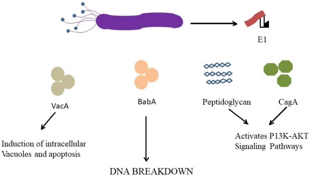
Carcinogenic effect of H. pylori through different mechanisms. Copyright Simona Cavalu et al. H. pylori infections and alternative treatment approaches. Copyright Simona Cavalu et al.
H. pylori is responsible for a chronic, transmissible, infectious disease and the increasing prevalence of antibiotic resistance has complicated the therapy. All therapies should assume the possibility of antimicrobial drug resistance.
In the context of increasing rates of antibiotic-resistant H. pylori strains, the risk factors and prevalence on global population, the aim of our work is to highlight the main drawbacks of currently used treatment regimens against H. pylori and at the same time, to emphasize the huge potential of natural alternatives, plants extracts and new formulation design and strategies to combat this pathogen. Special attention is also given to nanotechnological formulations, with huge potential for tissue microenvironment-responsive treatment. Copyright Simona Cavalu et al.
Nanotechnology-Based Approach against H. pylori Infections
Metallic NPs such as silver, gold, zinc or iron have been previously reported to possess the ability of killing a wide range of bacteria including H. pylori [153,154] by well-known underlying mechanisms involving oxidative stress, metal ion release and nonoxidative stress. A very low NPs concentration is necessary for bactericidal effect, and hence, it is difficult for the bacteria to develop resistance. Among different metallic NPs, AgNPs are convenient, especially the biologically derived ones, as the preparation methods demonstrated a controlled particle size, shape, and mono-dispersity, while reducing time of preparation, in the context of environmentally friendly approaches. Copyright Simona Cavalu et al.
Hydrogel original formulation. Copyright Simona Cavalu et al.
The preparation of gel formulation containing Trifolium pratense L. and Ocimum basilicum L. extract.
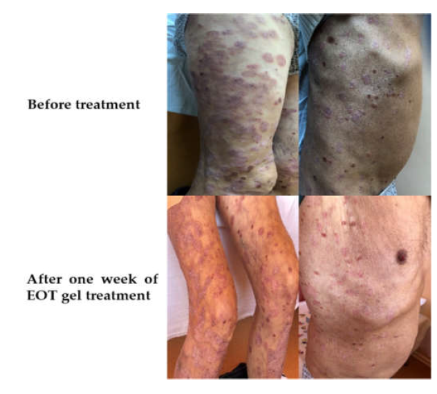
Spontaneous migration of dermal fibroblasts and evolution of “gap’’ closure in time: Copyright Simona Cavalu et al.Evolution of the migration of fibroblasts and “gap coverage” (which is similar to wound closure), with respect to the statistical factor “Sample”. Copyright Simona Cavalu et al.Animal model. Evolution in time (contraction) of the wound healing process in both groups (the control group and the EOT hydrogel-treated group). Copyright Simona Cavalu et al.Clinical aspects of Psoriasis vulgaris treated with gel formulation of Ocimum basilicum and Trifolium pratense extract mixture. Copyright Simona Cavalu et al.