Revisiting Therapeutic Strategies for H. pylori Treatment in the Context of Antibiotic Resistance: Focus on Alternative and Complementary Therapies

By I.A. Cardos, D. C. Zaha, R.K. Sindhu and Simona Cavalu

Copyright Simona Cavalu et al.
Carcinogenic effect of H. pylori through different mechanisms. Copyright Simona Cavalu et al.
Copyright Simona Cavalu et al.
H. pylori infections and alternative treatment approaches. Copyright Simona Cavalu et al.

H. pylori is responsible for a chronic, transmissible, infectious disease and the increasing prevalence of antibiotic resistance has complicated the therapy. All therapies should assume the possibility of antimicrobial drug resistance.

In the context of increasing rates of antibiotic-resistant H. pylori strains, the risk factors and prevalence on global population, the aim of our work is to highlight the main drawbacks of currently used treatment regimens against H. pylori and at the same time, to emphasize the huge potential of natural alternatives, plants extracts and new formulation design and strategies to combat this pathogen. Special attention is also given to nanotechnological formulations, with huge potential for tissue microenvironment-responsive treatment. Copyright Simona Cavalu et al.

Nanotechnology-Based Approach against H. pylori Infections

Metallic NPs such as silver, gold, zinc or iron have been previously reported to possess the ability of killing a wide range of bacteria including H. pylori [153,154] by well-known underlying mechanisms involving oxidative stress, metal ion release and nonoxidative stress. A very low NPs concentration is necessary for bactericidal effect, and hence, it is difficult for the bacteria to develop resistance. Among different metallic NPs, AgNPs are convenient, especially the biologically derived ones, as the preparation methods demonstrated a controlled particle size, shape, and mono-dispersity, while reducing time of preparation, in the context of environmentally friendly approaches. Copyright Simona Cavalu et al.

https://www.mdpi.com/1420-3049/26/19/6078

The power of plants. Evaluation of Wound Healing Potential of Novel Hydrogel Based on Ocimum basilicum and Trifolium pratense Extracts

By Simona Cavalu et al.

Copyright Simona Cavalu et al.
Hydrogel original formulation. Copyright Simona Cavalu et al.

The preparation of gel formulation containing Trifolium pratense L. and Ocimum basilicum L. extract.

Copyright Simona Cavalu et al.
Spontaneous migration of dermal fibroblasts and evolution of “gap’’ closure in time: Copyright Simona Cavalu et al.
Copyright Simona Cavalu et al.
Evolution of the migration of fibroblasts and “gap coverage” (which is similar to wound closure), with respect to the statistical factor “Sample”. Copyright Simona Cavalu et al.
Copyright Simona Cavalu et al.
Animal model. Evolution in time (contraction) of the wound healing process in both groups (the control group and the EOT hydrogel-treated group). Copyright Simona Cavalu et al.
Copyright Simona Cavalu et al.
Clinical aspects of Psoriasis vulgaris treated with gel formulation of Ocimum basilicum and
Trifolium pratense extract mixture. Copyright Simona Cavalu et al.

https://www.mdpi.com/2227-9717/9/11/2096

Electrochemical (Bio)Sensors with improved performances for Biomedical Applications

By L. Fritea, Simona Cavalu et al.

Metal Nanoparticles and Carbon-Based Nanomaterials for Improved Performances of Electrochemical (Bio)Sensors with Biomedical Applications

 A “real-time” biosensor includes a biological recognition receptor (such as an antibody, enzyme, nucleic acid or whole cell) and a transducer to convert the biological binding event to a detectable signal, which is read out indicating both the presence and concentration of the analyte molecule.In nano(bio)sensors, nanoparticles (NPs) are incorporated into the (bio)sensor design by attachment to the suitably modified platforms. For this purpose, metal nanoparticles have many advantageous properties making them useful in the transducer component of the (bio)sensors. Gold, silver and platinum NPs have been the most popular ones, each form of these metallic NPs exhibiting special surface and interface features, which significantly improve the biocompatibility and transduction of the (bio)sensor compared to the same process in the absence of these NPs. The main types of NPs used for electrochemical (bio)sensors design, especially screen-printed electrodes, with their specific medical application due to their improved analytical performances and miniaturized form is presented.

Copyright Simona Cavalu et al.
Schematic representation of main synthesis methods of metal NPs and carbon-based nanomaterials. Copyright Simona Cavalu et al.
Copyright Simona Cavalu et al.
Some examples of commercially available screen-printed electrodes (from different manufacturers: Copyright Simona Cavalu et al.

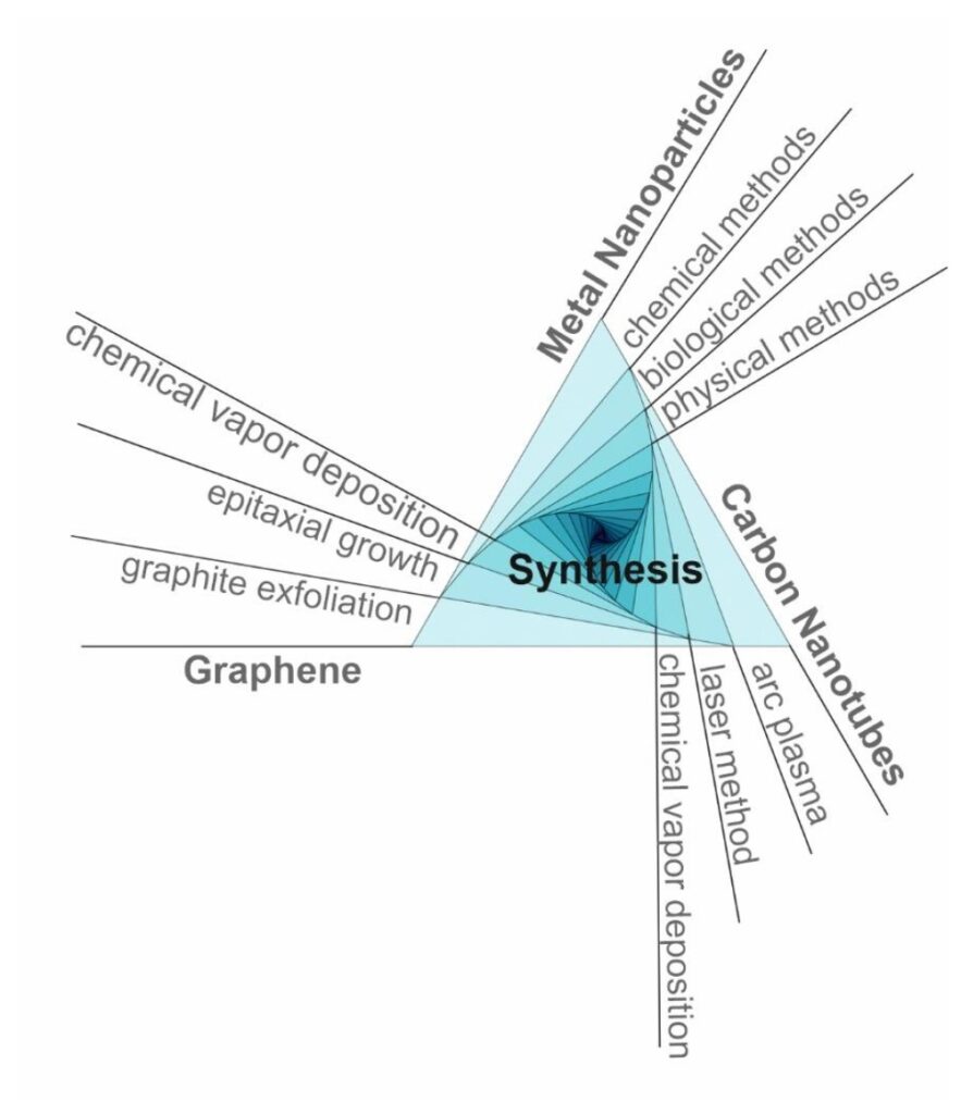
This comprehensive review is focused on the main types of metal NPs and carbon-based nanomaterials used for electrochemical (bio)sensors design, especially screen-printed electrodes, with their specific biomedical applications, improved analytical performances and miniaturized form.Nanotechnological approaches will extend the limits of currently employed (bio)sensors and, moreover, they will open a new window toward personalized medicine, offering new solutions to the main challenges in the diagnostic and therapeutic fields. Future research should focus on some improvements concerning the nanomaterials characteristics and the sensor design in order to enhance their performances with multi-disciplinary efforts. The real sample analysis with more enhanced sensitivity and selectivity is still a challenge for researchers aiming the validation of the electrochemical nano(bio)sensors in comparison with the traditional analytical procedures. The reproducibility is another key aspect which needs to be solved for large-scale production of electrochemical sensors and their introduction on commercial market. The miniaturized, portable or wearable sensors which can perform on-site and real-time analysis will gain tremendous importance at the commercial level, with a huge impact on the health system.

The full text of this paper is available at

https://www.mdpi.com/1996-1944/14/21/6319/htm

Liposomes for oral delivery of Berberine

By H.I. Kutbi, H.Z. Asfour, A. K. Kammoun, A. Sirwi, H. A. Gad and Simona Cavalu

Optimization of Hyaluronate-Based Liposomes to Augment the Oral Delivery and the Bioavailability of Berberine

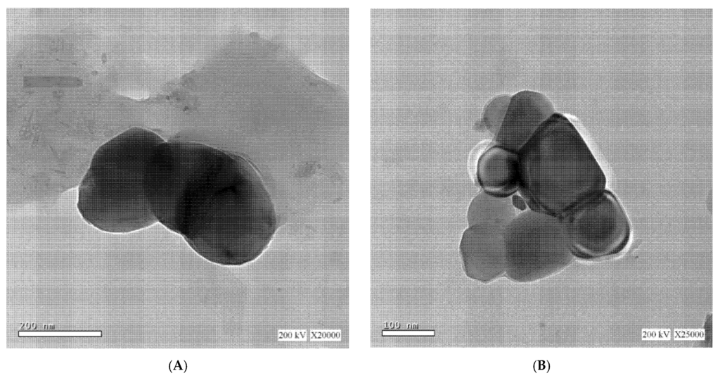
To improve Brb permeability and bioavailability, this study presents a newly developed formulation, namely Brb hyaluronate-based liposomes, prepared by using film hydration method and characterized by dynamic light scattering measurements, entrapment efficiency percentage (EE%), transmission electron microscope (TEM), in vitro drug release and physical stability. Results of pharmacokinetics studies indicated the potential of the liposomal formulation to increase the oral bioavailability of Brb and to accelerate its entry into the bloodstream. The obtained results are accredited to the lipophilic nature of the prepared system, resembling the structural features of bio-membrane, in addition to their small size that enhances intestinal penetration. 

Berberine hyaluronate based liposomes. Copyright H. A. Gad and Simona Cavalu
Transmission electron microscope of berberine hyaluronate based liposomes, showing the nearly spherical shape (A) and the multi-lamellar structure of the vesicles bilayer (B) of the prepared liposomes. Copyright H. A. Gad and Simona Cavalu
The effect of the independent variables on liposomes particle size and entrapment efficiency percent. Copyright H. A. Gad and Simona Cavalu
Three-dimensional (3D) response surface plots showing the effect of the independent variables on liposomes particle size and entrapment efficiency percent: A—total lipid amount (mg), B—berberine (mg), C—hyaluronic acid (mg). Copyright H. A. Gad and Simona Cavalu
Plasma concentration profiles of berberine in rats after oral administrations. Copyright H.A. Gad and Simona Cavalu
Plasma concentration profiles of berberine in rats after oral administrations of 50 mg/kg of berberine in various formulations. Each point represents mean ± standard deviation (N = 6). Copyright H.A. Gad and Simona Cavalu

Different formulation variables (lipid, drug and hyaluronic acid amounts) have a significant effect on the physicochemical characteristics of the prepared system using film hydration method. The presence of hyaluronic acid as a main component in liposomes preparation was able to slow berberine diffusion from the vesicles.Oral administration of Brb hyaluronate-based liposomes to rats could improve lipophilicity and bioavailability of the investigated system compared to Brb solution and Brb liposomes prepared without hyaluronic acid. Copyright H.A. Gad and Simona Cavalu

The full text of this paper is available at

https://www.mdpi.com/1996-1944/14/19/5759/htm

Carbon Nanotubes for Improved Performances of Endodontic Sealer

By Simona Cavalu et al.

In order to overcome the limitations of current endodontic sealers, especially against resistant bacteria, recent developments in the field of nanotechnology have proved the necessity to reconsider the composition and physico-chemical properties of classical sealers. Nanoparticles with their unique features in terms of small size and high specific surface area, are the best choice for incorporation of antiseptic agents and effective delivery. Multi-walled carbon nanotubes (MWCNTs) encapsulating chlorhexidine (CHX) and colloidal silver nanoparticles (AgNPs) were prepared and incorporated into commercial sealer and investigated in terms of bonding performance to dentin and effectiveness against E. faecalisS. aureus and Candida albicans, which are responsible for the majority of the failures in endodontic treatments. In this context, the challenges related to the long-term biological effects of CHX/AgNPs loaded MWCNTs are discussed.

Development of “smart” endodontic therapeutic agents 

SEM morphological details of CNTs. Copyright Simona Cavalu
SEM morphological details of CNTs as received from the manufacturer, before (a) and after (b) loading with the mixture CHX/AgNPs (High magnification, 100,000×).
Copyright Simona Cavalu et al.
Colloidal CNTs in distilled water (left) compared to colloidal mixture CHX/AgNPs. Copyright Simona Cavalu
SEM morphological details of CNTs as received from the manufacturer, before (a) and after (b) loading with the mixture CHX/AgNPs (High magnification, 100,000×); (c) photographic image of colloidal CNTs in distilled water (left) compared to colloidal mixture CHX/AgNPs (right). Copyright Simona Cavalu et al.
SEM  details of commercial  and modified root canal sealer. Copyright Simona Cavalu
Ultrastructure details of commercial (a) and modified root canal sealer (c) along with the corresponding EDX spectrum (b,d). Copyright Simona Cavalu et al.
Load–displacement curves and Young modulus calculation. Copyright Simona Cavalu.
Load–displacement curves (a) recorded on the surface of commercial (black) and modified sealer (red) and the corresponding Young modulus calculation (b). Copyright Simona Cavalu et al.
TGA and DTG thermograms of the modified and commercial sealers.
Copyright Simona Cavalu et al.
SEM images of interfacial adaptation between sealer and root canal dentine. Copyright Simona Cavalu
SEM images (a,c) of interfacial adaptation between sealer and root canal dentine (polished specimens) along with the corresponding EDX spectra (b,d): (a) neat sealer;
(c) CNTs/CHX/AgNPs modified sealer. The transversal section was performed in the middle zone of the root. Copyright Simona Cavalu et al.
Antimicrobial and antifungal effect of different combinations and the mixture CNTs/AgNPs/CHX2%
Antimicrobial and antifungal effect of different combinations and the mixture CNTs/AgNPs/CHX2% against the tested strains. Data are expressed as average value ± standard deviation of triplicate samples (statistical significance * p < 0.05).
Copyright Simona Cavalu

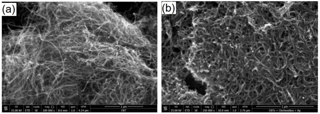
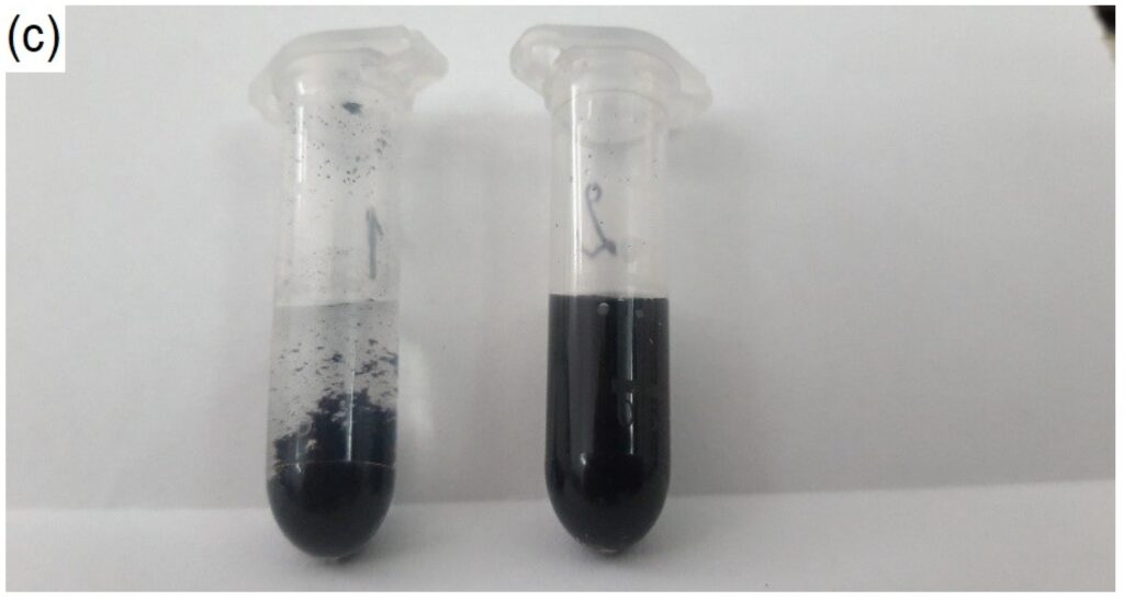
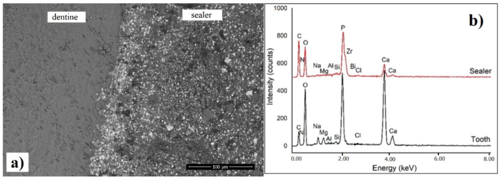
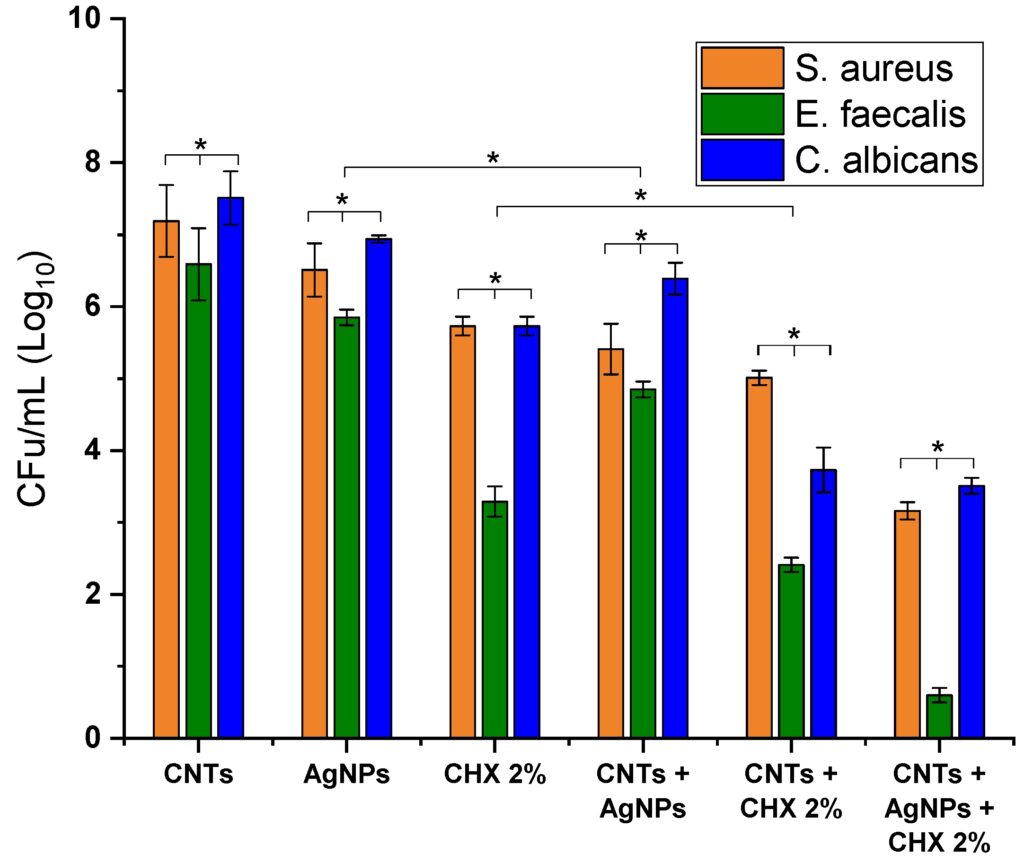
Our original approach, in the context of new generation sealers expecting to have a long-lasting antimicrobial effect, was to demonstrate that the antimicrobial effect of the mixture CNTs/AgNPs/CXH 2% incorporated in commercial sealer, was preserved long enough to efficiently inhibit Gram-positive germs, with excellent results towards E. faecalis in a concentration of 1 mg/mL. The antibacterial and antifungal assay clearly demonstrated a synergic effect of AgNPs, CHX 2% and CNTs with excellent results towards E. faecalis, which is responsible for the primary etiologic factors in pulp and periapical lesions.

The full text of this paper is available at

https://www.mdpi.com/1996-1944/14/15/4248/htm

Functional food based on elemental selenium NPs

By S. I. Vicas, Simona Cavalu at al.

Nano Selenium—Enriched Probiotics as Functional Food
Products against Cadmium Liver Toxicity

The main goal of our work was to develop a functional food based on elemental selenium nanoparticles (SeNPs) obtained by green synthesis using Lactobacillus casei and to validate their ability to annihilate the hepatic toxic effects induced by cadmium.A functional food that includes both probiotic bacteria and elemental SeNPs could be successfully used to annihilate Cd-induced liver toxicity, and to improve both nutritional values and health benefits.

TEM micrograph of SeNPs synthesized using L. casei and NaHSeO3 as a reducing agent. Copyright Simona Cavalu et al.
TEM micrograph of SeNPs synthesized using L. casei and NaHSeO3 as a reducing agent; (b) energy dispersive X-ray analysis; and (c) particles size distribution confirmed by DLS. Copyright Simona Cavalu et al.

We proposed investigating for the first time the protective effect of SeNPs and lacto-SeNPs (LSeNPs) administered orally to mice for 30 days in different concentrations (0.1, 0.2 and 0.4 mg/kg b.w.), against the toxic effects exerted by cadmium at the hepatic level. Blood biochemical parameters (transaminases, bilirubin, gamma glutamyl transferase), antioxidant enzymes (catalase and glutathione peroxidase), the antioxidant capacity of plasma along with the histology, immunohistochemistry for mitochondrial apoptosis markers (bcl-2bax) and gene expression of hepatic inflammatory markers (NF-ĸB, TNFα, IL-6) were analyzed in terms of the comparative evaluation of the dose-dependent protective activity of SeNPs and LSeNPs against cadmium intoxication.

The histopathological sections of mice livers of the experimental groups.
Copyright Simona Cavalu et al.
Hepatic immunohistochemical expression of bcl-2 (A) and bax (B). Copyright Simona Cavalu et al.

Co-administration of Cd with both forms of SeNPs significantly decreased the gene expression of liver inflammatory markers, with the best effects for LSeNPs. A functional food that includes both probiotic bacteria and elemental SeNPs could be successfully used to annihilate Cd-induced liver toxicity, and to improve both nutritional values and health benefits. In this way, a possible new technology is provided for the food industry, the production of yogurt enriched with selenium nanoparticles produced by lactic acid bacteria with protective effects against heavy metals.

The full text of the paper can be accessed at:

https://www.mdpi.com/1996-1944/14/9/2257/htm

Amniotic membrane for ophthalmologic surgery

By Simona Cavalu et al.

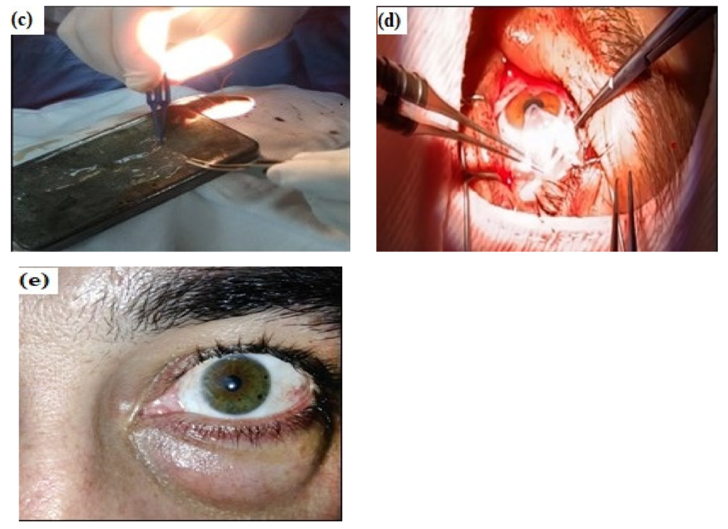
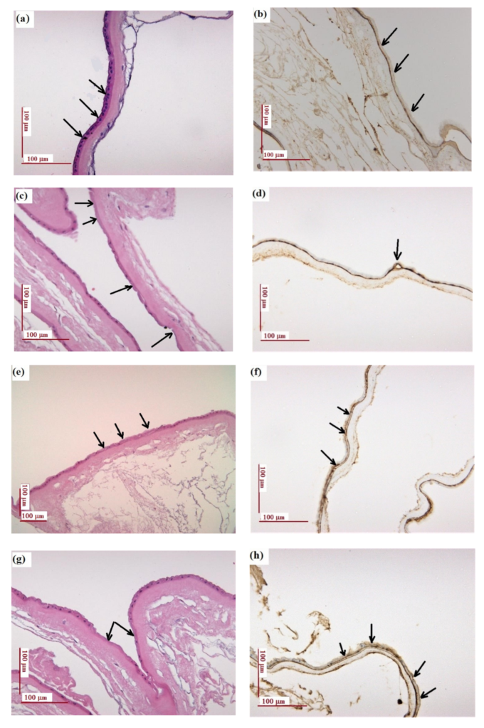
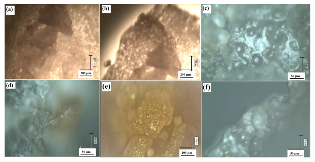
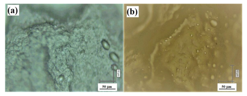
Nano-Scale Modifications of Amniotic Membrane Induced by UV and Antibiotic Treatment: Histological, AFM and FTIR Spectroscopy Evidence

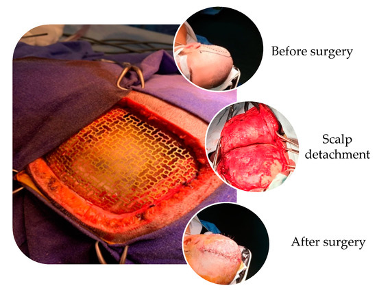
The efficiency of amniotic membrane (AM) transplantation in different types of ocular surface disorders is due to its outstanding properties such as antifibrotic, antibacterial, anti-inflammatory and antiangiogenic, working as a versatile scaffold to promote corneal tissue epithelialization.

Recurrent pterygium excision. Copyright Simona Cavalu et al.
Amniotic membrane used in ophthalmologic surgery to cover the remaining tissue defect due to the grade 3 recurrent pterygium excision: (a) grade 3 recurrent pterygium, preoperative appearance; (b) intraoperative aspects (the same patient); (c) intraoperative preparation of the amniotic membrane fragment to be applied on the remaining defect following recurrent pterygium excision; (d) intraoperative appearance—application of the AM fragment on the remaining defect and 10.0 thread suturing to the bulbar conjunctiva of the fragment; (e) the aspect of the patient’s cornea four weeks after surgery . Copyright Simona Cavalu et al.
 Histological and immunohistochemical examination of amniotic membrane. Copyright Simona Cavalu et al.
 Histological and immunohistochemical examination: (a,b) natural amniotic membrane (AMN); (c,d) amniotic membrane exposed to UV for 1 h (AUV); (e,f) amniotic membrane treated with gentamicin (40 mg/mL) (AG40); (g,h) amniotic membrane treated with gentamicin 80 mg/mL (AG0); (i,j) amniotic membrane treated with gentamicin 40 mg/mL and exposed to UV for 1 h (AGUV). Left panel: H&E staining; right panel: immunohistochemistry staining of collagen IV (antibody clone CIV 22). Scale: 100 μm. Copyright Simona Cavalu et al.

The full text of the paper is available at:

https://www.mdpi.com/1996-1944/14/4/863/htm

Rapid Screening of Retrieved Knee Prosthesis Components by Confocal Raman Micro-Spectroscopy

By Simona Cavalu et al.

Bilateral knee radiography—anteroposterior view, demonstrating advanced degenerative changes (gonarthrosis stadium IV) at the level of the right knee (white arrows), secondary to the malunion of tibial plateau fracture; (b) lateral view, showing depression of the lateral tibial plateau (white arrow). Copyright Simona Cavalu et al.
(c) anteroposterior and later view, eight years after surgery; both views highlight the loosening of the tibial component, minimal osteolysis of the medial tibial plateau (anteroposterior view) and minimal osteolysis around the distal region of the tibial implant (lateral view); (d) anteroposterior and lateral view after revision of the total knee arthroplasty using modular tibial implant with short stem and two screws for re-insertion of the patellar tendon (after tibial tuberosity osteotomy). Copyright Simona Cavalu et al.
Photographic images of retrieved tibial components of knee prosthesis. (a) Assembled tibial components; (b) titanium component and synovial fluid collected from the stem (white arrow); (c) separate metallic and ultra-high-molecular-weight polyethylene (UHMWPE) component, showing surface delamination (white arrow); (d) acrylic cement detached from the metallic component. Copyright Simona Cavalu et al.
Micro-Raman spectra recorded on the surface of tibial metallic component (titanium). (a) As supplied from the producer; (b) retrieved prosthetic component along with the corresponding microscopic image (inset). Scale bar 50 µm. Copyright Simona Cavalu et al.
Micro-Raman spectra acquired from the surface of plastic (UHMWPE) component. Copyright Simona Cavalu et al.
Comparison of Micro-Raman spectra acquired from the surface of plastic (UHMWPE) component of a new (green spectra) and retrieved (red) knee prosthesis, showing the vibrational changes (band shifts, relative intensity change) during 10 years aging in human physiological environment. The details of significant intervals are shown in (a) 960–1200 cm−1, (b) 1280–1310 cm−1, (c) 1400–1500 cm−1 and (d) 2820–2960 cm−1 spectral ranges. Spectra acquired under similar excitation and optical collection conditions (excitation: 785, 30 mW, 10 s acquisition, 1 accumulation, 20× objective). Multiple spectra collected from single points of old plastic are shown. Black spectra refer to color spotted plastic supposed to be due to organic micro-deposits, however, the decreased polyethylene signal only was observed. Copyright Simona Cavalu et al.
Micrographs recorded in real time on the surface of UHMWPE component of knee prosthesis during Confocal Raman measurements. Copyright Simona Cavalu et al.
Micro-Raman spectra collected in the low (A) and high wavenumber range (B) from the surface of acrylic cement debris detached from the metallic stem, characteristic for acrylic-bone interface; and (C,D) synovial fluid spectra from light (a) and dark (b) spotted points, accumulated in the stem of the tibial component. Copyright Simona Cavalu et al.
Micrographs corresponding to multiple acquisition performed on different sites of the surface of acrylic cement debris. The images shows details of the acrylic cement surface in contact with the (a,b) metallic component and with (cf) the biologic tissue.
Copyright Simona Cavalu et al.
Micrographs corresponding to multiple acquisition performed on different sites on the surface of synovial fluid, showing embedded lipid droplets: (a) center, (b) edge.
Copyright Simona Cavalu et al.

Retrieved knee prosthesis

Sursa MDPI