Nano-Scale Modifications of Amniotic Membrane Induced by UV and Antibiotic Treatment: Histological, AFM and FTIR Spectroscopy Evidence
The efficiency of amniotic membrane (AM) transplantation in different types of ocular surface disorders is due to its outstanding properties such as antifibrotic, antibacterial, anti-inflammatory and antiangiogenic, working as a versatile scaffold to promote corneal tissue epithelialization.
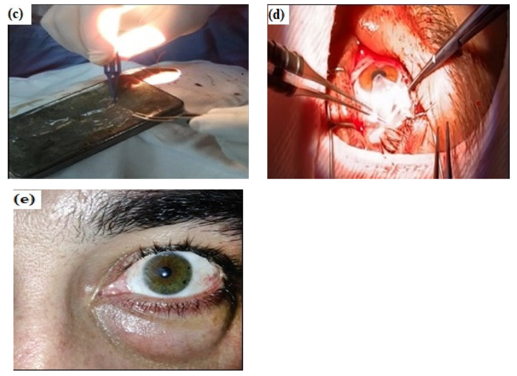
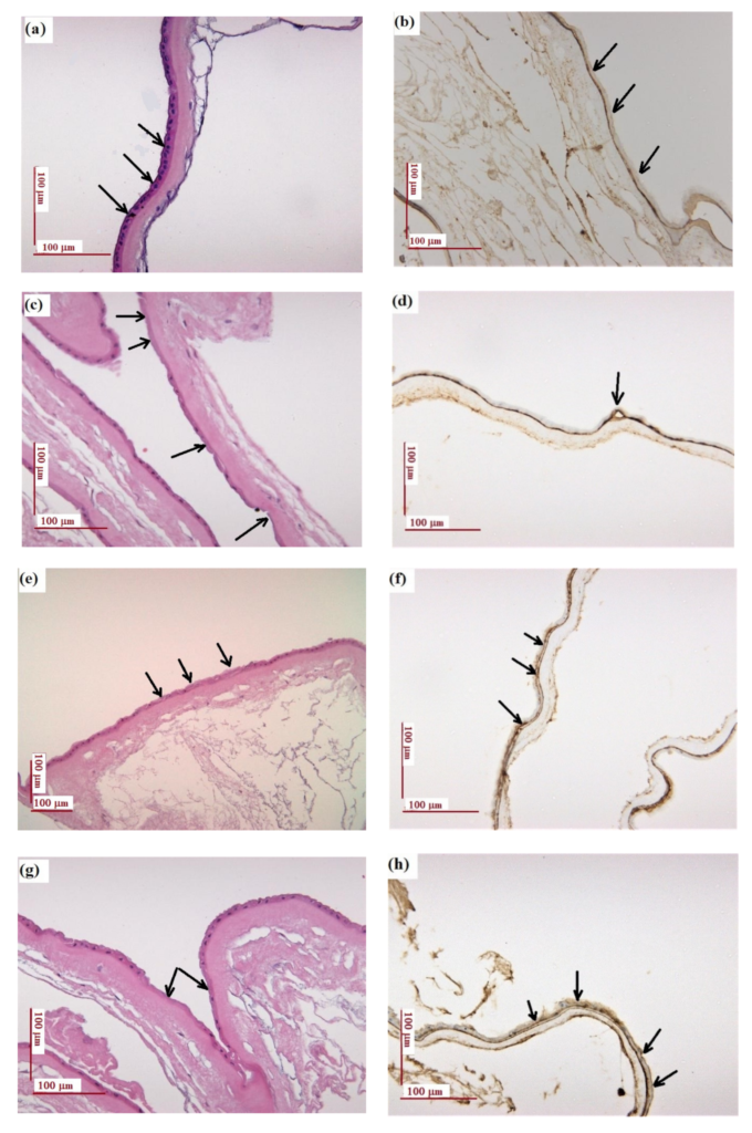
Amniotic membrane used in ophthalmologic surgery to cover the remaining tissue defect due to the grade 3 recurrent pterygium excision: (a) grade 3 recurrent pterygium, preoperative appearance; (b) intraoperative aspects (the same patient); (c) intraoperative preparation of the amniotic membrane fragment to be applied on the remaining defect following recurrent pterygium excision; (d) intraoperative appearance—application of the AM fragment on the remaining defect and 10.0 thread suturing to the bulbar conjunctiva of the fragment; (e) the aspect of the patient’s cornea four weeks after surgery . Copyright Simona Cavalu et al. Histological and immunohistochemical examination: (a,b) natural amniotic membrane (AMN); (c,d) amniotic membrane exposed to UV for 1 h (AUV); (e,f) amniotic membrane treated with gentamicin (40 mg/mL) (AG40); (g,h) amniotic membrane treated with gentamicin 80 mg/mL (AG0); (i,j) amniotic membrane treated with gentamicin 40 mg/mL and exposed to UV for 1 h (AGUV). Left panel: H&E staining; right panel: immunohistochemistry staining of collagen IV (antibody clone CIV 22). Scale: 100 μm. Copyright Simona Cavalu et al.