by Adela Ciorba, Simona Cavalu et al.
Full text at http://dx.doi.org/10.21614/chirurgia.3160
by A.L. Ciorba, A. Teusdea, G. Roiu and Simona Cavalu
The aim of this study was to evaluate the influence of ultrasounds used in phacoemulsification during cataract surgery on the corneal structure and morphology in patients over 65 years. We compared the outcomes of phacoemulsification techniques in terms of corneal cell morphology in
77 patients over 65 years old and 43 patients under 65 years old. Corneal cell density, central corneal thickness and hexagonality were measured preoperatively and post-surgery (at 1 and 4 weeks) by specular microscopy. The effect of gender, axial length and anterior chamber depth on the parameters of corneal endothelium were evaluated. In both groups, a progressive decrease in endothelial cells was observed, starting from the first week post-surgery until the fourth postoperative week. The central
corneal thickness increased in both groups with maximum values at the first week postoperatively, while their initial values were restored in the fourth week post-surgery, with no statistical difference between groups. Statistically significant differences were noticed in terms of cell hexagonality in the group over 65, showing smaller hexagonality at all preoperative and postoperative time points compared to group under 65. Our result highlights the importance of routine specular microscopy
performed before surgery, regardless the age of the patients, with caution and careful attention to the phaco power intensity, ultrasound energy consumption and intraoperative manipulation of instruments, as well as proper use of viscoelastic substances to reduce corneal endothelium damage, especially in elderly patients. Copyright Simona Cavalu et al.
Cataract is considered a multifactorial eye disease, due to the opacification of the lens, that leads to visual impairment when is located in the visual axis. The main cause for cataract development is ageing together with oxidative stress. Risk factors associated with cataract are known to be
educational and income status, smoking, diabetes, ultraviolet radiation, body mass index, estrogen replacement therapy, drug use (non-steroidal antiinflammatory drugs), traumatic injuries, chemicals and local diseases like uveitis or retinal detachment. The surgical technique, the anterior
chamber depth and the use of ophthalmic viscoelastic devices are other factors that can influence the damage degree of the endothelium, as well as the type of IOLs implanted (toric versus non-toric), especially in patients with corneal degeneration or dystrophies. Studies report endothelial cell loss rates from 4% to 25% after phacoemulsification. In this context, the aim of our study was to investigate the main outcomes of cataract surgery in terms of corneal cell density (CD), central corneal thickness (CCT) and hexagonality (HEX) of the endothelial cells in patients over 65 years of age compared to those under 65. Copyright Simona Cavalu et al.
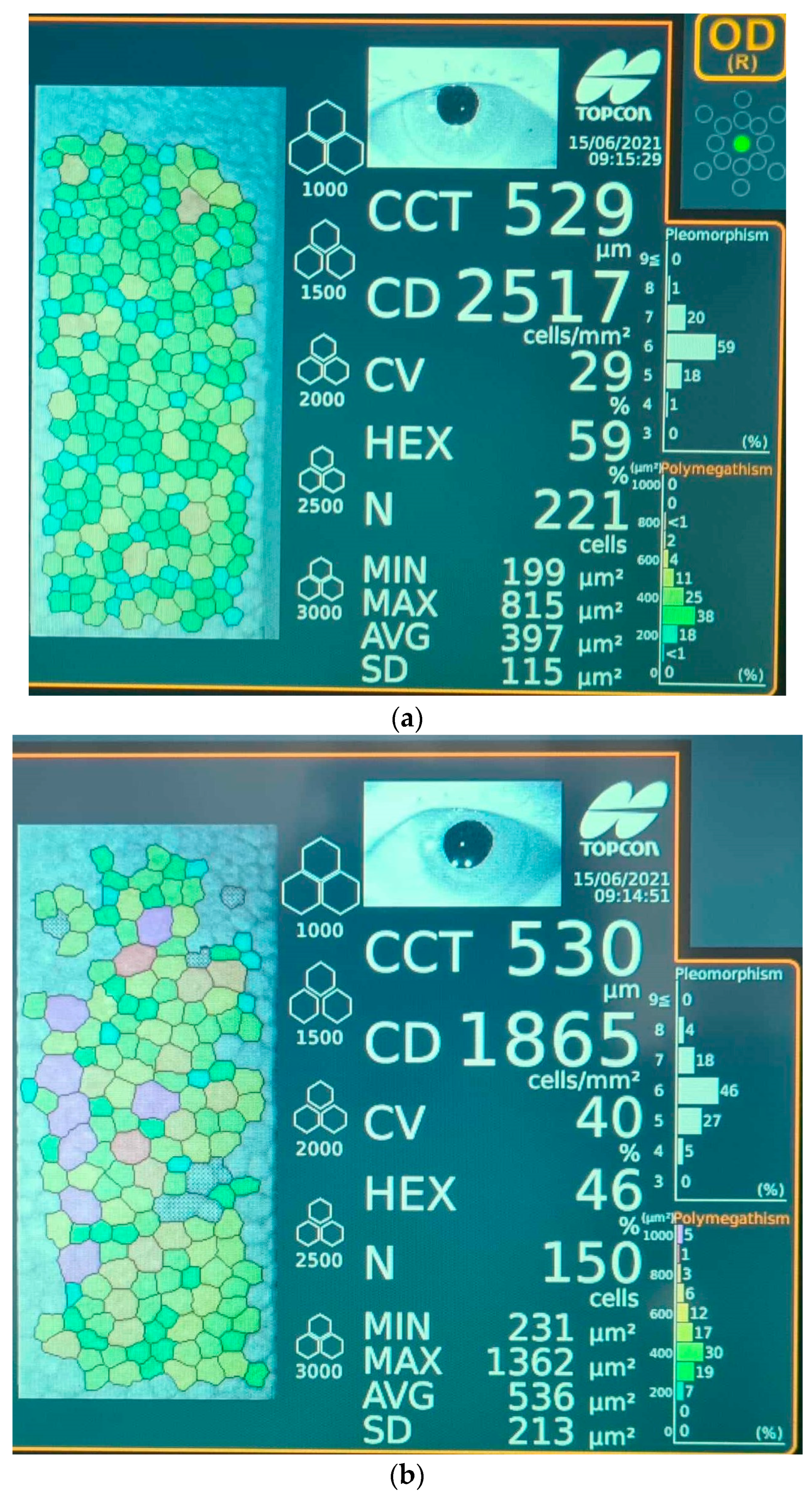

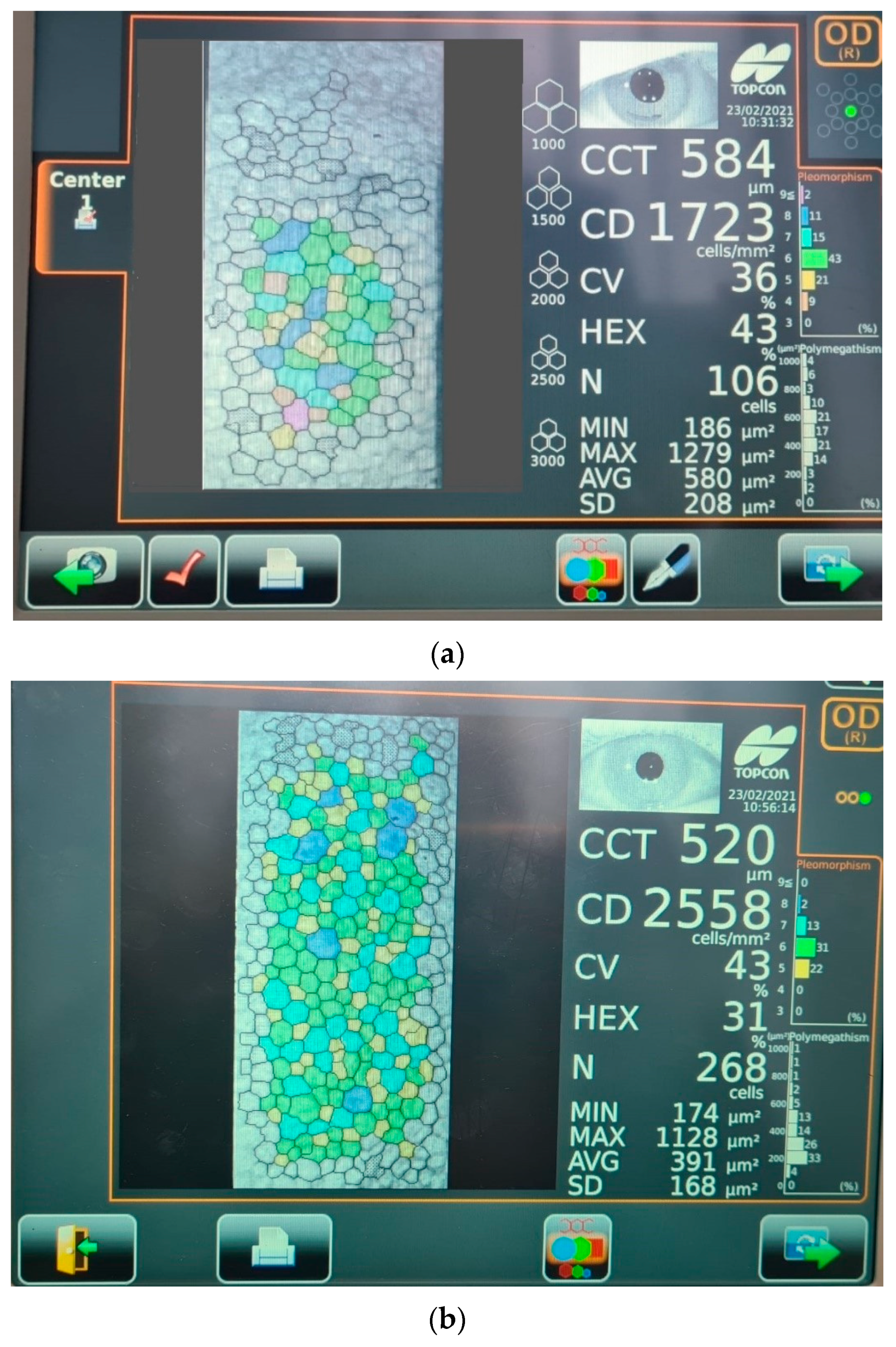
Endothelial Cell Density (CD)
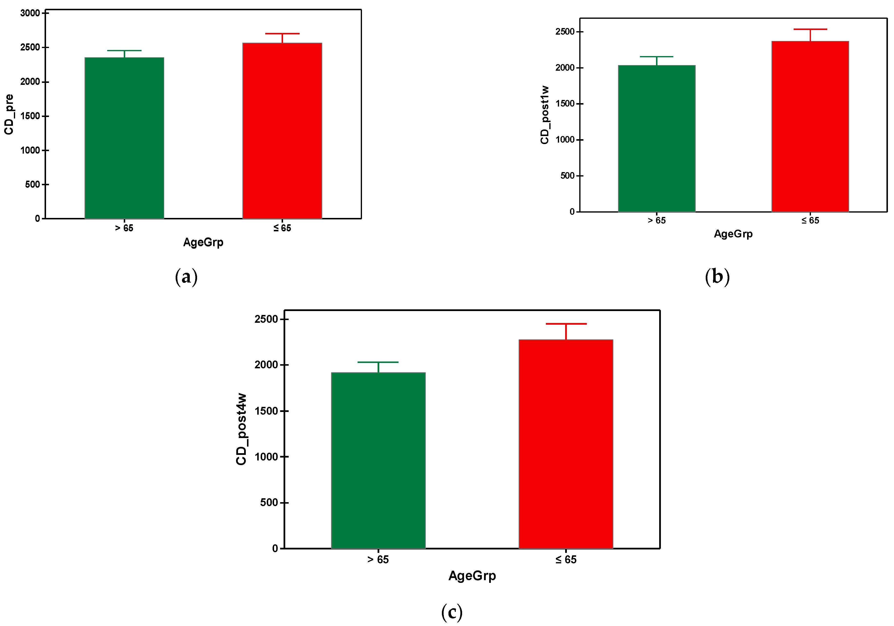


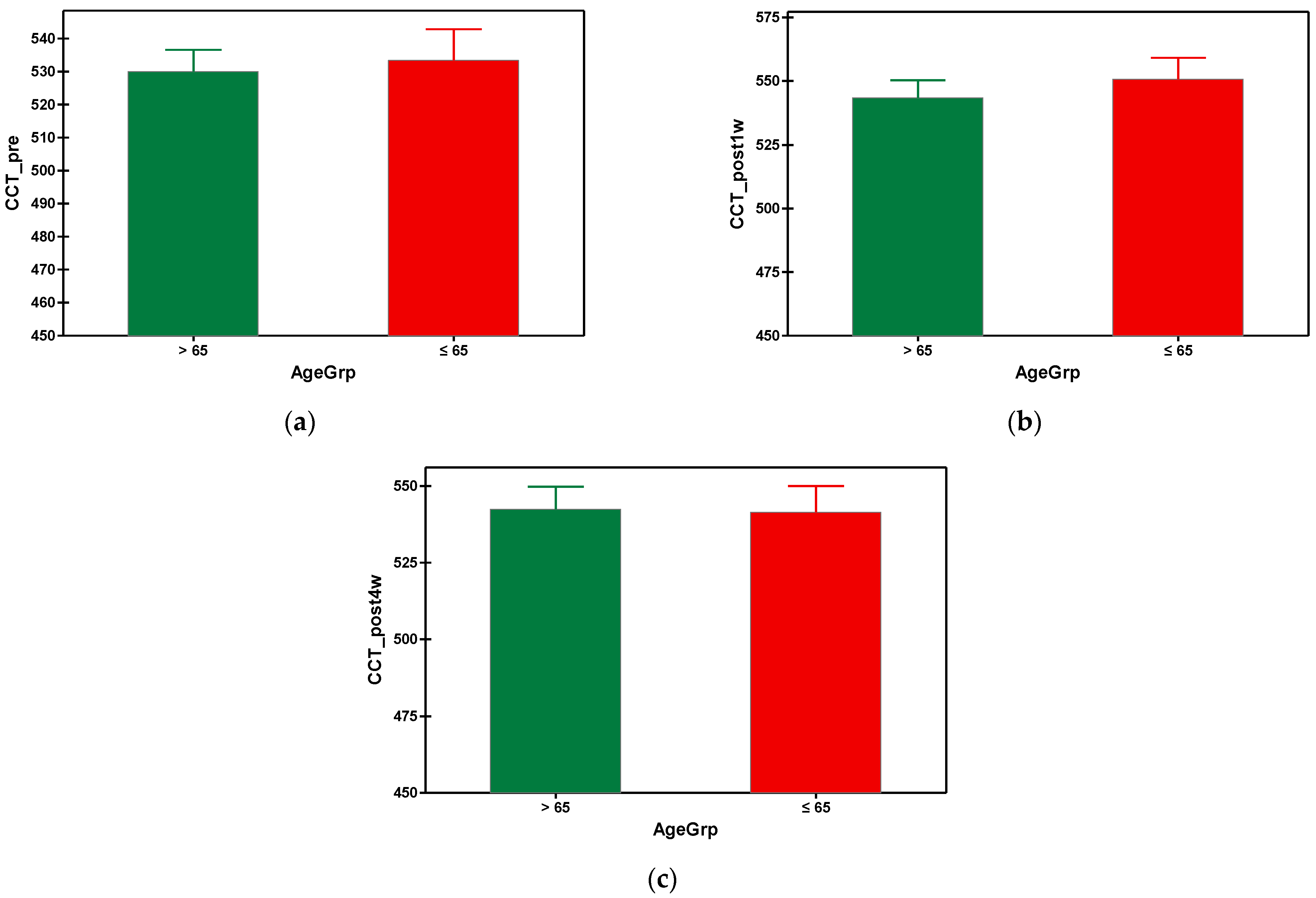
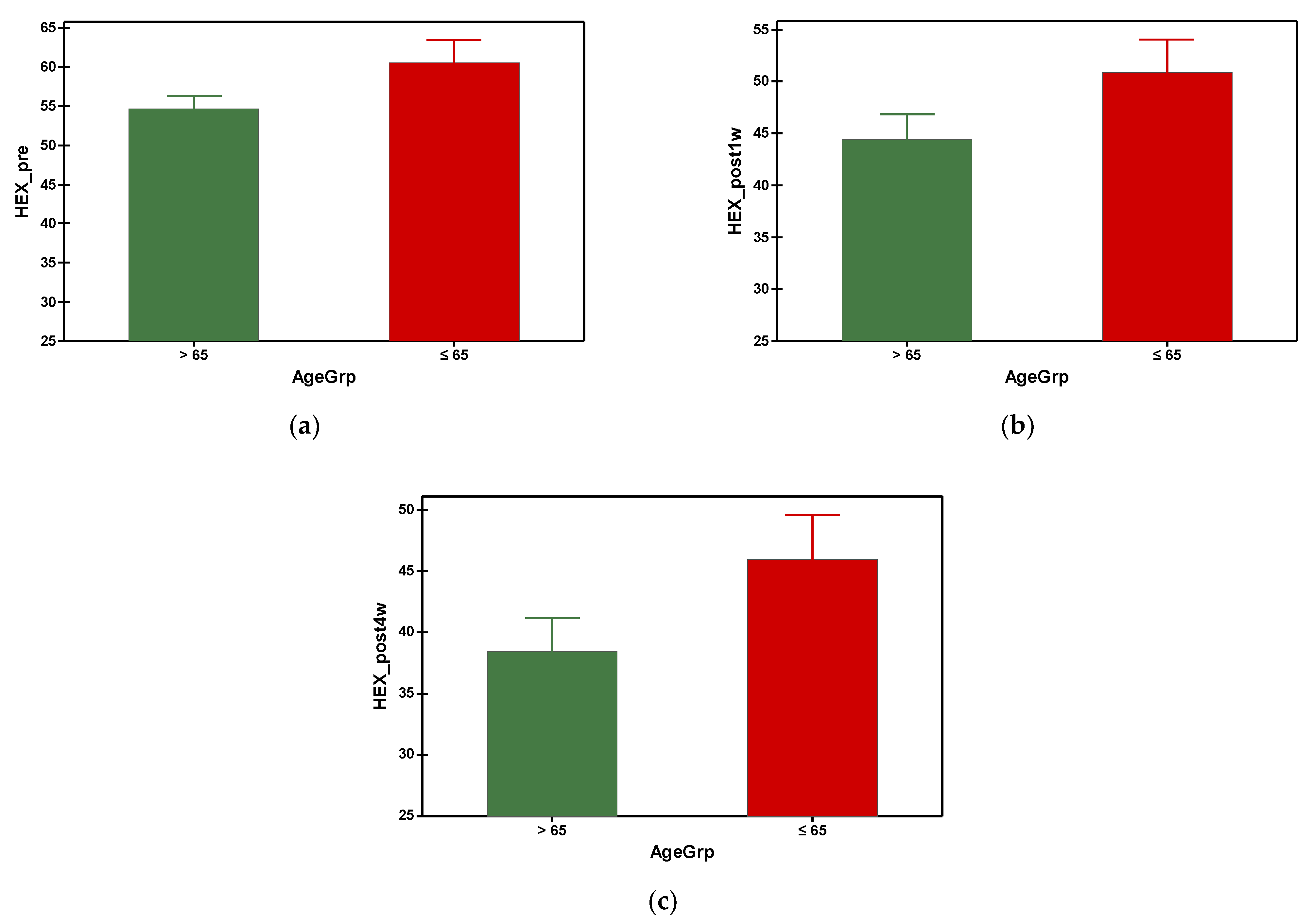

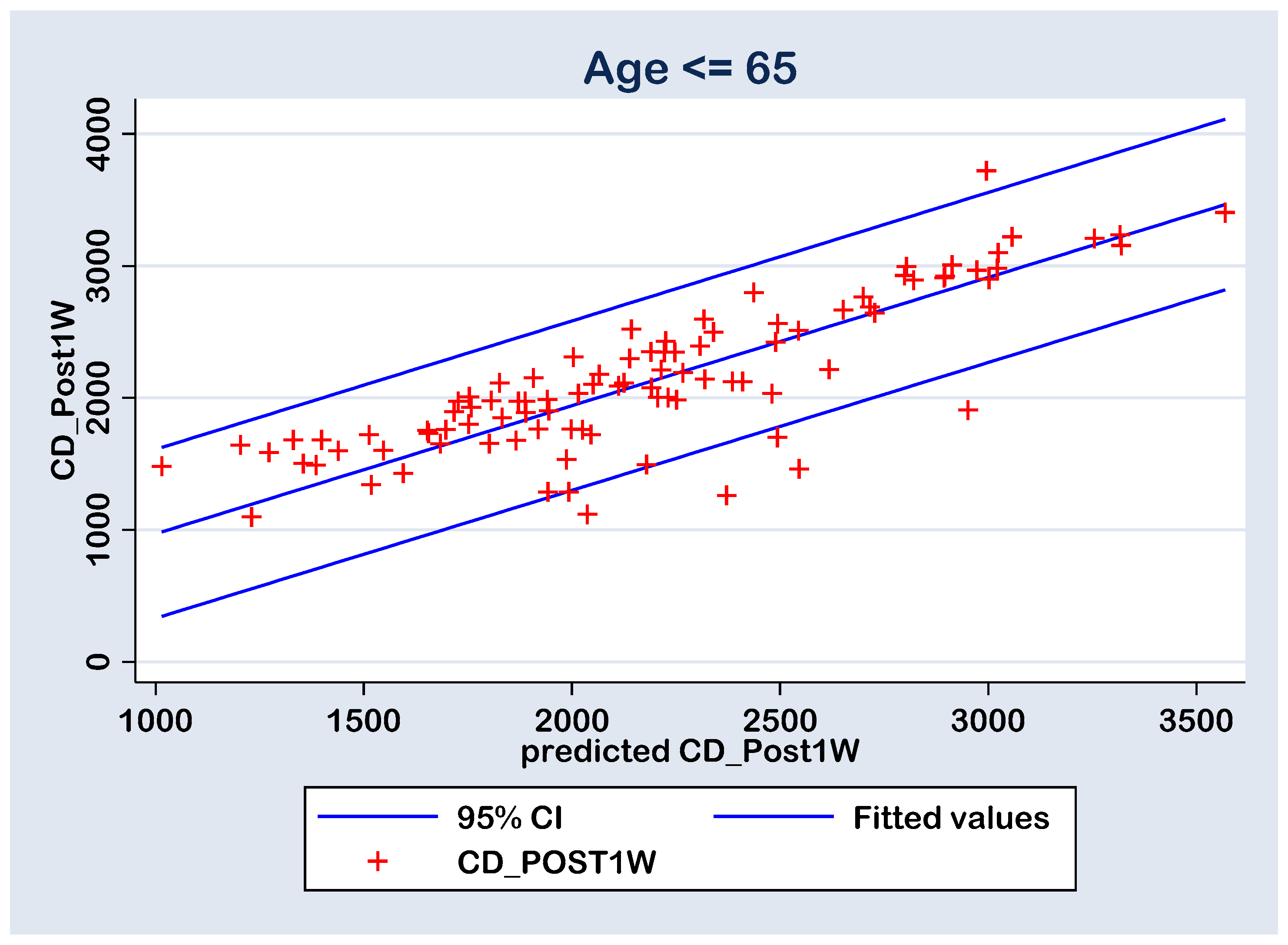
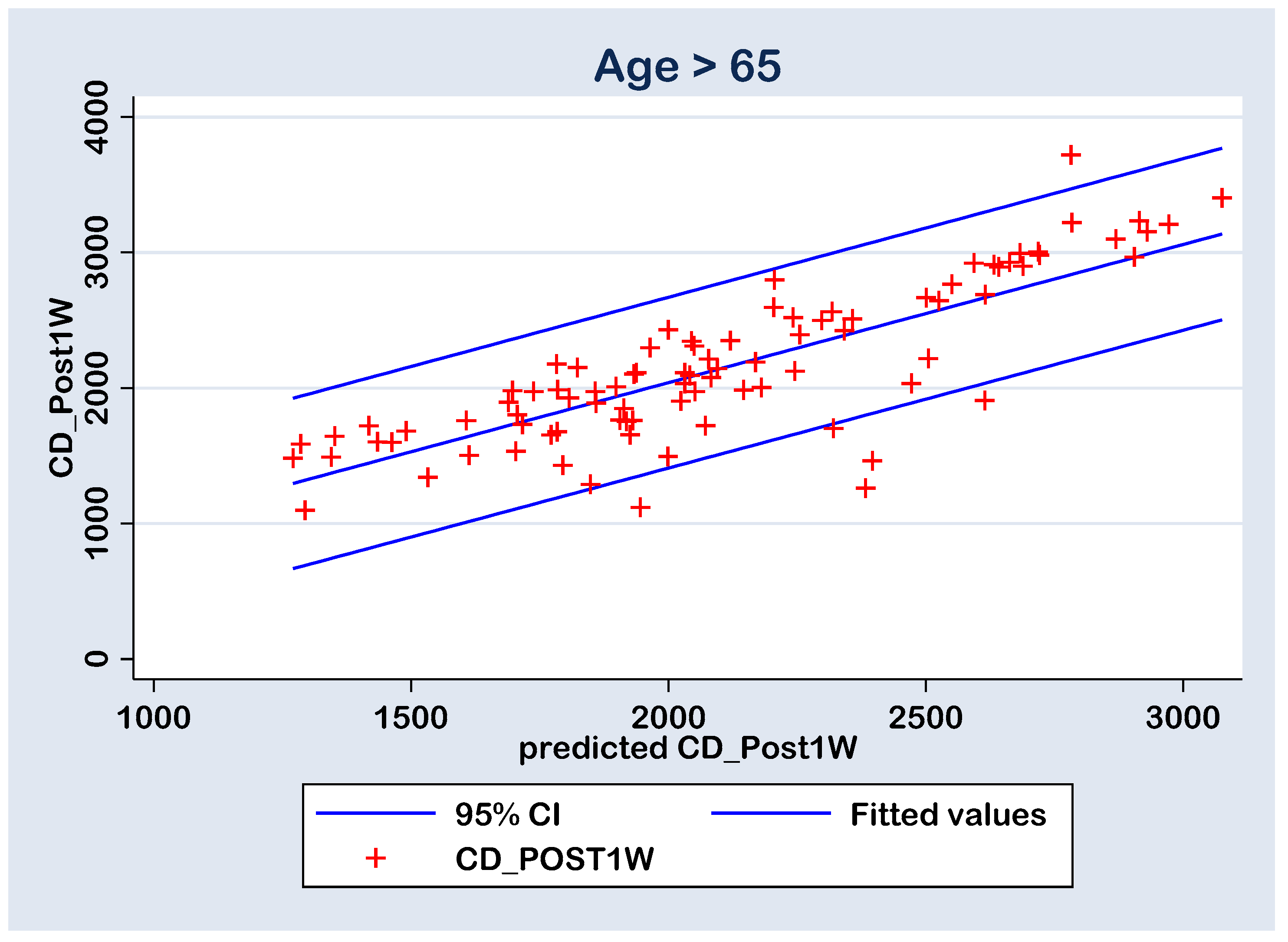
Our study evaluated the influence of phacoemulsification cataract surgery technique on corneal cell density, central corneal thickness and hexagonality preoperatively and postoperatively at 1 and 4 weeks using the specular microscope. The effect of gender, axial length and anterior chamber depth on the parameters of corneal endothelium in the two groups, under and over 65 years of age were evaluated. There were significant differences between the preoperative and postoperative cell density values in both groups, with a progressive loss of cells until the fourth week of evaluation. A significant increase in the CCT was observed in both groups after the first week, while the initial values were restored to almost normal by the fourth week post-surgery, with no statistical difference between group A and B. The cell hexagonality suffered a progressive drop in both groups, with significantly lower HEX values at all preoperative and postoperative times in group A (>65 y). Although the cell density loss was significant in both groups, but more advanced in older patients, accompanied with increased central corneal thickness and reduced values of hexagonality, we had no cases of endothelial decompensation secondary to surgical trauma, characterized by epithelial and subepithelial bullae and stromal edema (also known as bullous keratopathy) that is directly responsible for vision loss following cataract surgery. This outcome can be associated to a good preoperative assessment of the patient, together with the surgeon’s experience and the application of viscoelastic substances with protective role on the corneal endothelium.
Phacoemulsification can and truly does improve the visual function of all patients, under and over 65 years old with senile or presenile cataracts, giving them fully independence for daily activities, improving their life quality. The use of specular microscopy in all patients undergoing phacoemulsification and intraoperative proper use of viscoelastic substances for endothelium protection, as well as making adjustments of intraoperative parameters used for nucleus removal according to preoperative cell density values and caution in terms of phaco power intensity and ultrasound energy, for a successful cataract surgery are all strongly recommended. Copyright Simona Cavalu et al.
The full text of this article at https://www.mdpi.com/2308-3417/9/3/77

By Adela Ciorba, Amir M. Abdelhamid, G. Roiu, S. Saber and Simona Cavalu
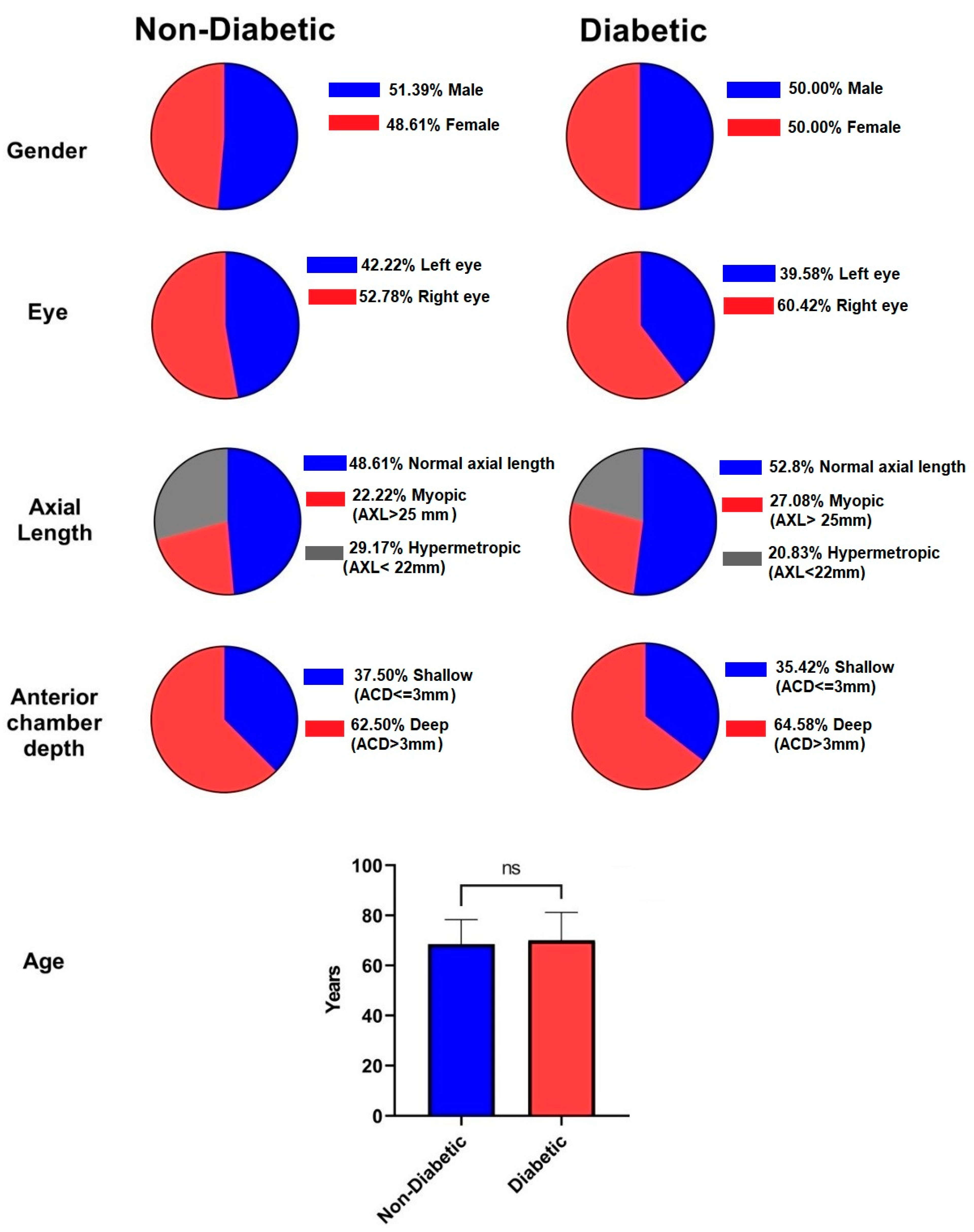
The aim of this study was to evaluate the influence of phacoemulsification cataract surgery on the state of the corneal endothelium in diabetic versus non-diabetic patients. We compared the corneal cell morphology in 48 diabetics with good glycemic control and 72 non-diabetic patients before and after uneventful phacoemulsification. Corneal cell density, central corneal thickness, and hexagonality were measured preoperatively and post-surgery (at 1 and 4 weeks) by specular microscopy. The effect of age, gender, axial length, and anterior chamber depth on the parameters of
the corneal endothelium were evaluated. Copyright Simona Cavalu et al.
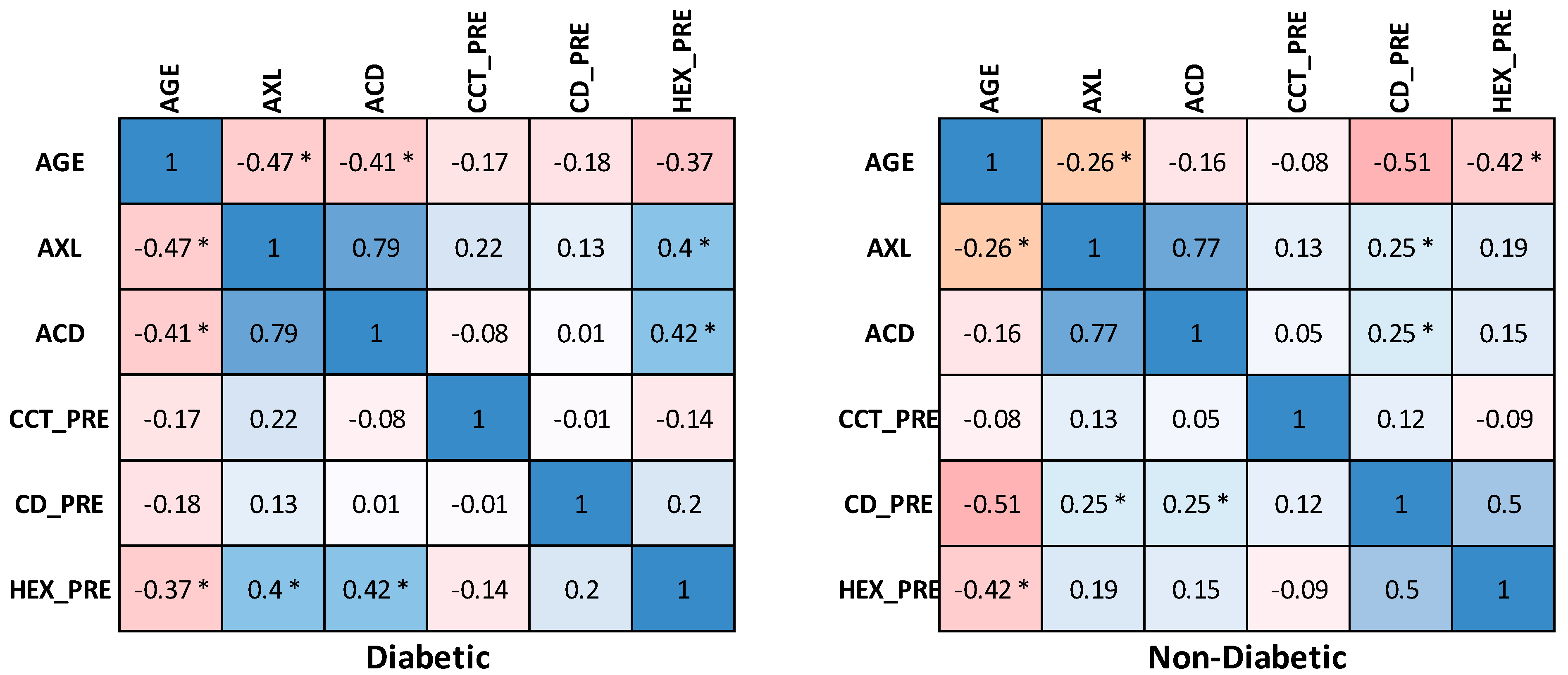
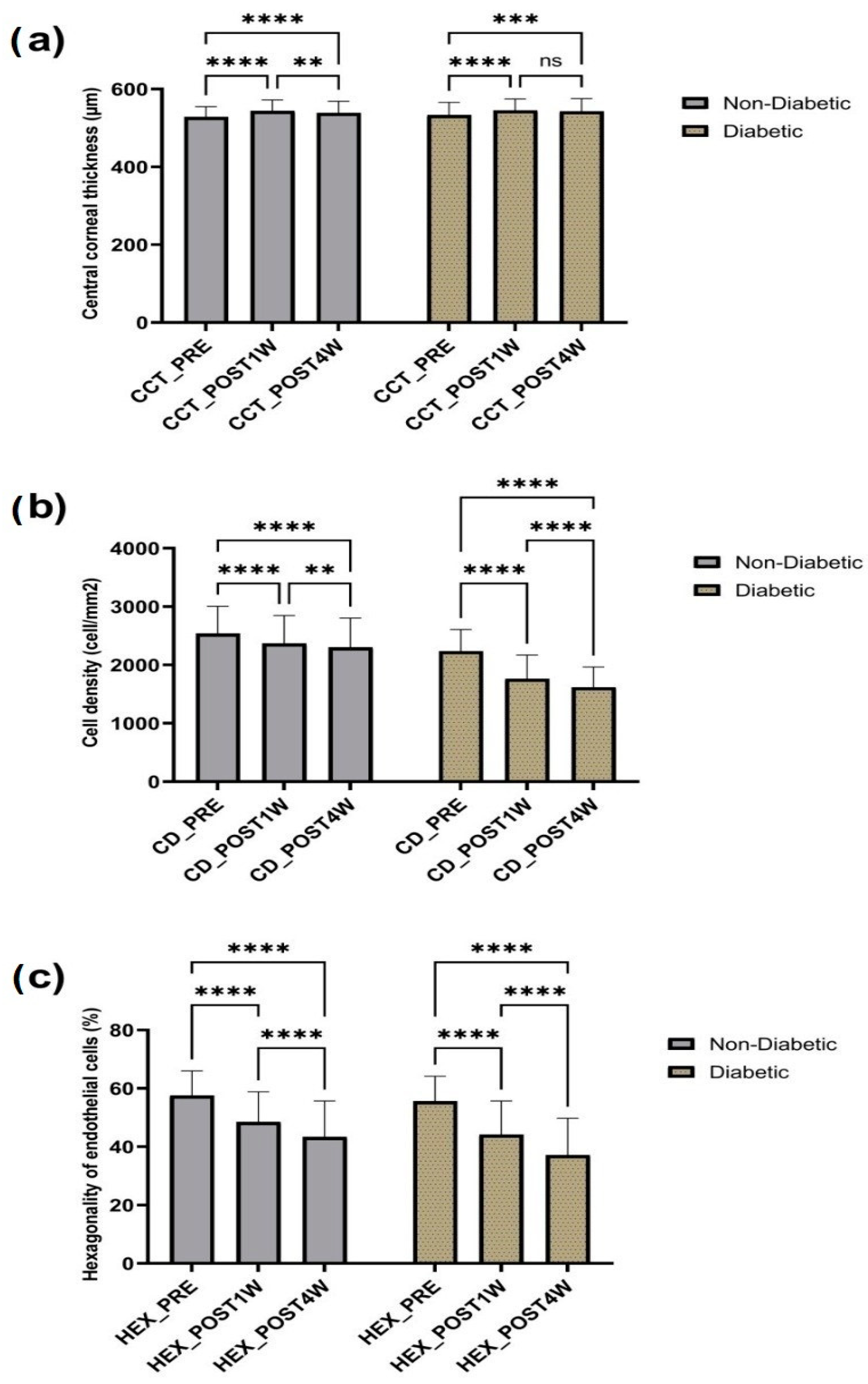
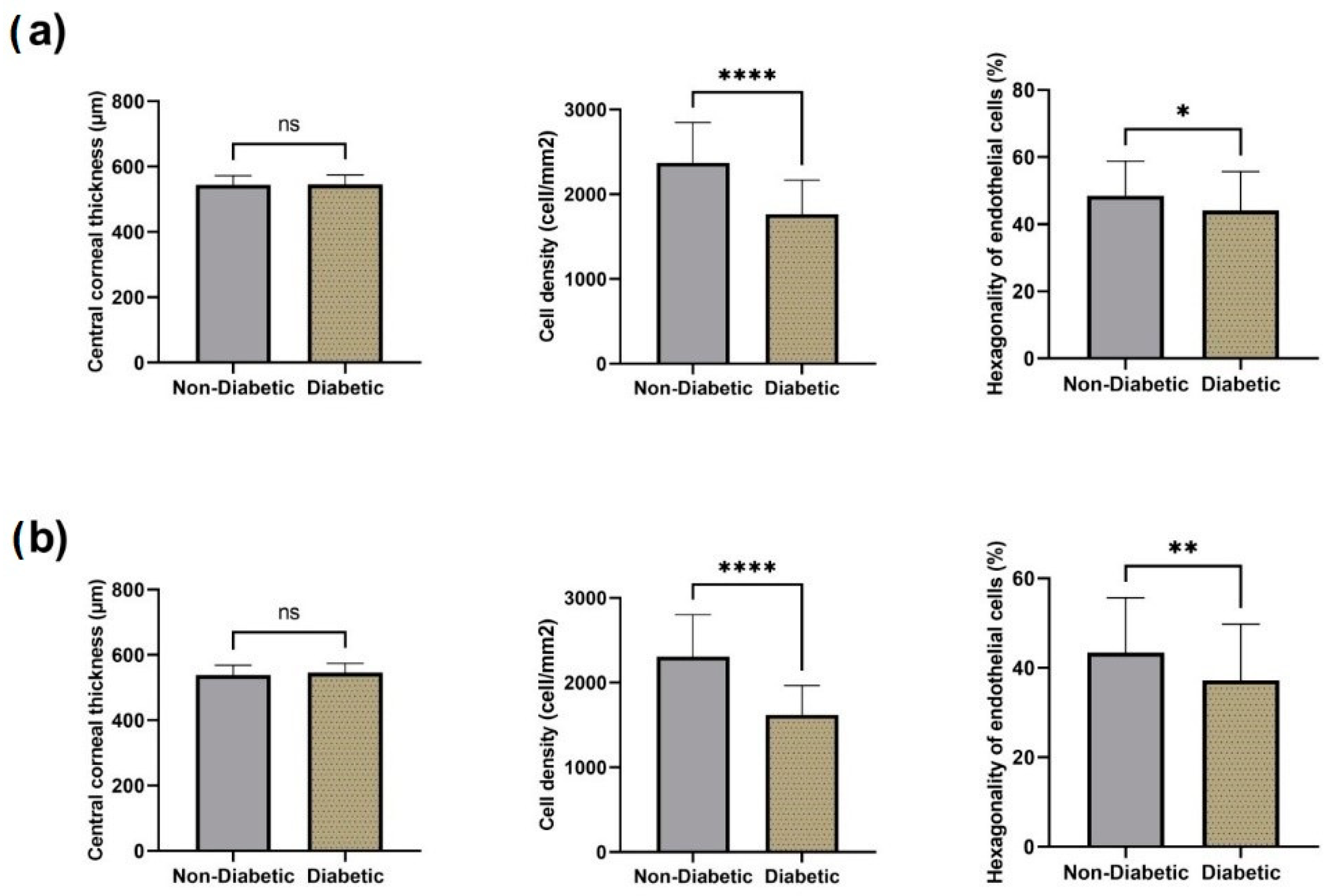
We noticed significant differences between pre-surgical and postoperative CD values in both diabetic and non-diabetic patients. Despite good glycemic control, diabetic patients had more pronounced morphological abnormalities compared to those of non-diabetics, but visual outcomes after phacoemulsification with IOL implantation were similar in both groups. A drop in the postoperative endothelial density was noted after the first week, in both groups. A significant increase in central corneal thickness was also noted in both groups one week after phacoemulsification, but there was no statistical significance after 4 weeks in the diabetic group. In terms of cell hexagonality, statistically significant differences were noted after 4 weeks in both groups.A major finding in our study is that, although an advanced loss of CD was noted, along with an increased CCT and a reduction of hexagonality (especially in the diabetic group), there were no cases of postoperative bullous keratopathy, probably due to several factors, such as surgeon’s experience and the use of viscoelastic substances with a protective role, as well as a careful preoperative evaluation and a good glycemic index (HbA1c < 7%).We strongly recommend routine specular microscopy and HbA1c evaluation before all cataract surgeries. Regarding intraoperative precautions, a high level of monitoring is necessary in terms of pacho power intensity and ultrasound energy, along with a proper application of the dispersive viscoelastic substances to reduce the risk of endothelial damage for a successful surgical procedure.
Copyright Simona Cavalu et al.
Full text here https://www.mdpi.com/2075-4418/13/6/1115