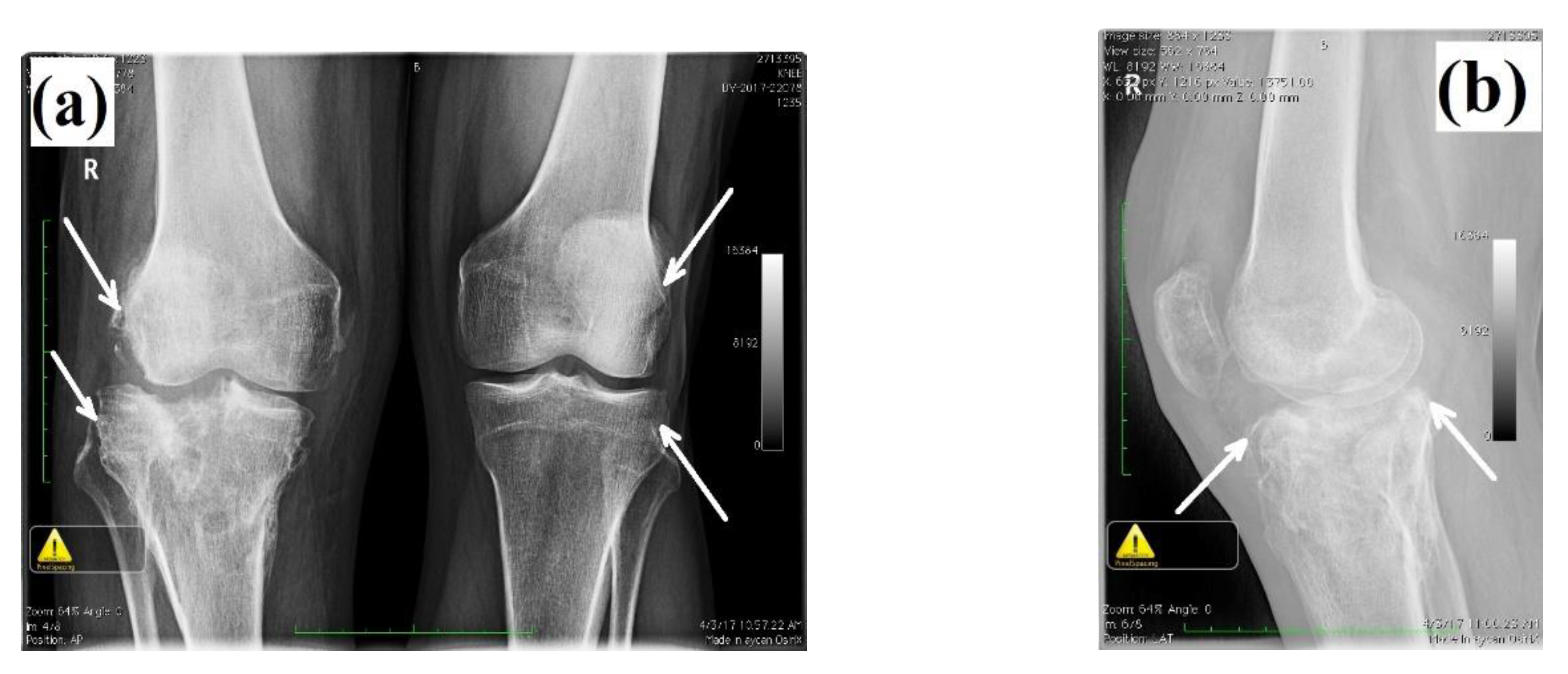
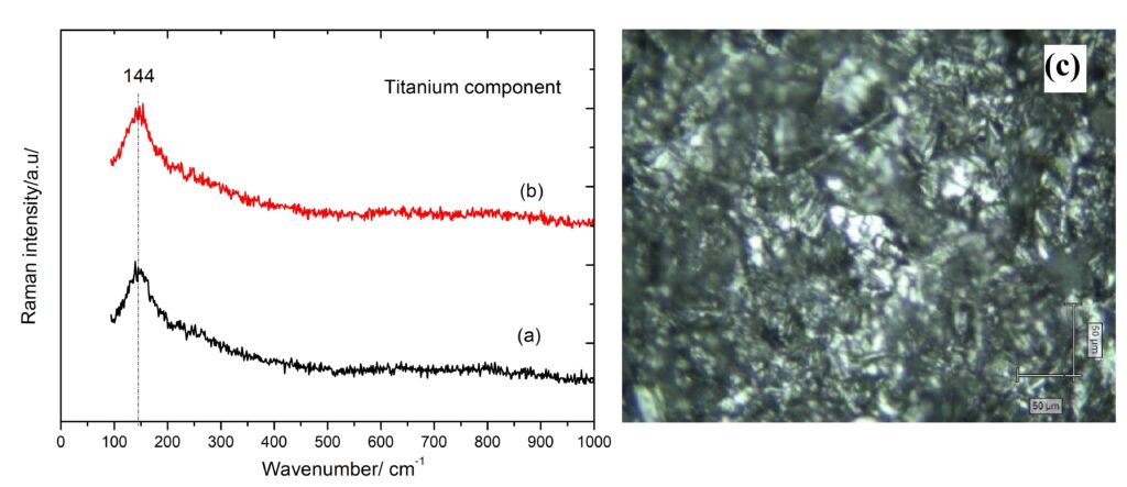
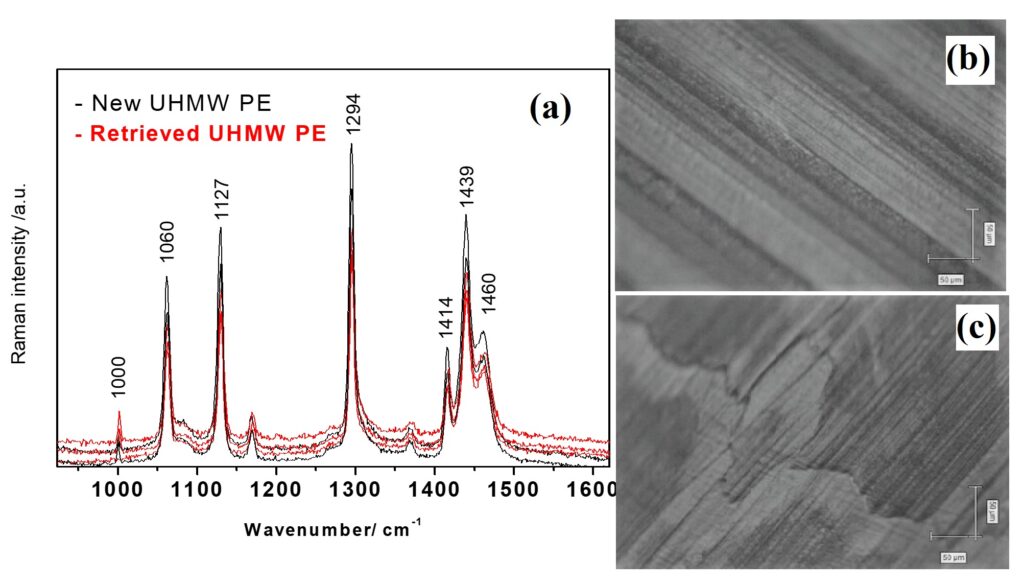
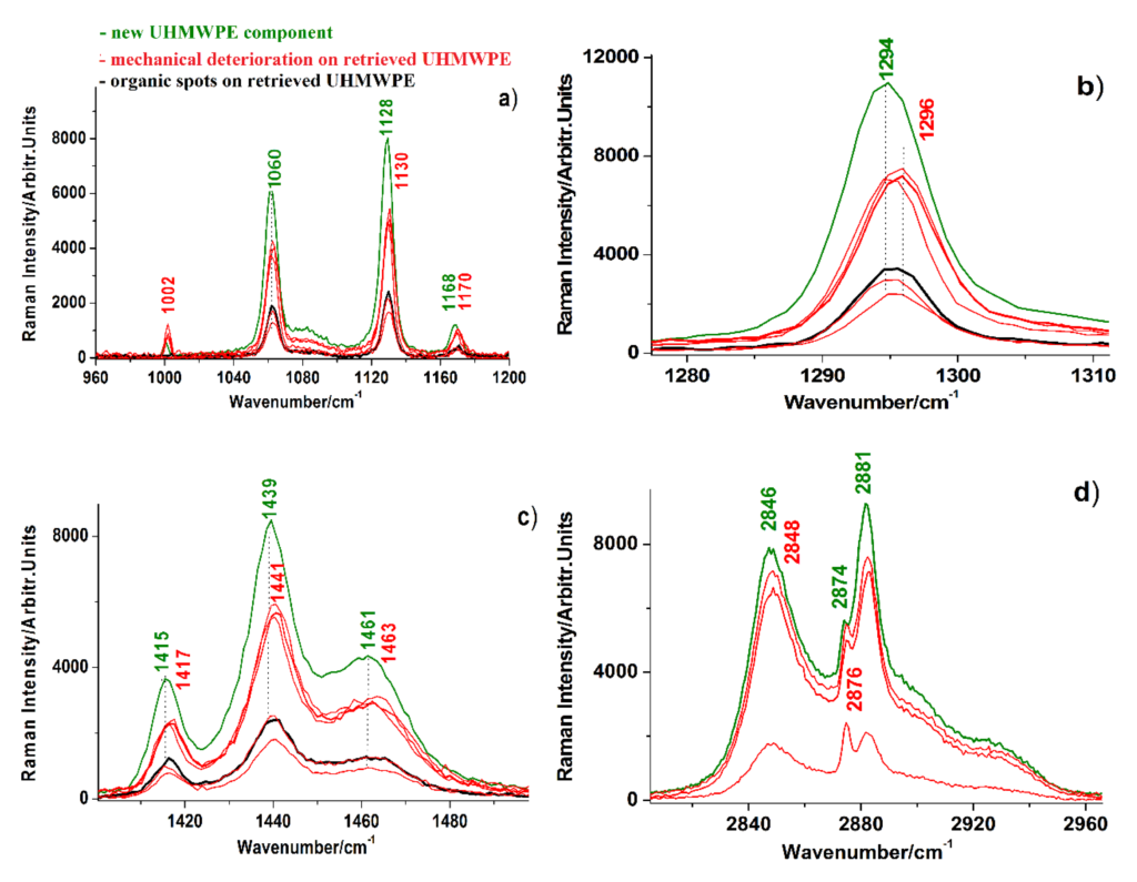
Bilateral knee radiography—anteroposterior view, demonstrating advanced degenerative changes (gonarthrosis stadium IV) at the level of the right knee (white arrows), secondary to the malunion of tibial plateau fracture; (b) lateral view, showing depression of the lateral tibial plateau (white arrow). Copyright Simona Cavalu et al.(c) anteroposterior and later view, eight years after surgery; both views highlight the loosening of the tibial component, minimal osteolysis of the medial tibial plateau (anteroposterior view) and minimal osteolysis around the distal region of the tibial implant (lateral view); (d) anteroposterior and lateral view after revision of the total knee arthroplasty using modular tibial implant with short stem and two screws for re-insertion of the patellar tendon (after tibial tuberosity osteotomy). Copyright Simona Cavalu et al.Photographic images of retrieved tibial components of knee prosthesis. (a) Assembled tibial components; (b) titanium component and synovial fluid collected from the stem (white arrow); (c) separate metallic and ultra-high-molecular-weight polyethylene (UHMWPE) component, showing surface delamination (white arrow); (d) acrylic cement detached from the metallic component. Copyright Simona Cavalu et al.Micro-Raman spectra recorded on the surface of tibial metallic component (titanium). (a) As supplied from the producer; (b) retrieved prosthetic component along with the corresponding microscopic image (inset). Scale bar 50 µm. Copyright Simona Cavalu et al.Micro-Raman spectra acquired from the surface of plastic (UHMWPE) component. Copyright Simona Cavalu et al.Comparison of Micro-Raman spectra acquired from the surface of plastic (UHMWPE) component of a new (green spectra) and retrieved (red) knee prosthesis, showing the vibrational changes (band shifts, relative intensity change) during 10 years aging in human physiological environment. The details of significant intervals are shown in (a) 960–1200 cm−1, (b) 1280–1310 cm−1, (c) 1400–1500 cm−1 and (d) 2820–2960 cm−1 spectral ranges. Spectra acquired under similar excitation and optical collection conditions (excitation: 785, 30 mW, 10 s acquisition, 1 accumulation, 20× objective). Multiple spectra collected from single points of old plastic are shown. Black spectra refer to color spotted plastic supposed to be due to organic micro-deposits, however, the decreased polyethylene signal only was observed. Copyright Simona Cavalu et al.Micrographs recorded in real time on the surface of UHMWPE component of knee prosthesis during Confocal Raman measurements. Copyright Simona Cavalu et al.Micro-Raman spectra collected in the low (A) and high wavenumber range (B) from the surface of acrylic cement debris detached from the metallic stem, characteristic for acrylic-bone interface; and (C,D) synovial fluid spectra from light (a) and dark (b) spotted points, accumulated in the stem of the tibial component. Copyright Simona Cavalu et al.Micrographs corresponding to multiple acquisition performed on different sites of the surface of acrylic cement debris. The images shows details of the acrylic cement surface in contact with the (a,b) metallic component and with (c–f) the biologic tissue. Copyright Simona Cavalu et al.Micrographs corresponding to multiple acquisition performed on different sites on the surface of synovial fluid, showing embedded lipid droplets: (a) center, (b) edge. Copyright Simona Cavalu et al.